Wear resistance of dental temporary restorations manufactured using different additive manufacturing technologies
Abstract
Recently, the digital industry has established itself as a major technology in the field of dentistry, while additive manufacturing (AM), commonly known as 3D printing. Temporary dental restorations are being produced using various additive manufacturing technologies. Also, changes in eating habits expose these temporary restorations to various acidity. Thus, this study was to investigate that wear resistance of dental temporary restorations manufactured using different additive manufacturing technologies according to the acidity of artificial saliva. A total of 120 rectangular parallelepipeds specimens were prepared with three different types of printing method used for the dental resin crown production: Stereo Lithography Apparatus (SLA), Digital Light Processing (DLP), Fused Deposition Modelling (FDM). The antagonists were made of zirconia and cobalt–chrome alloy. Each specimen was then loaded at 5 kg for 20,000 cycle chewing simulations with 5 mm vertical descending movement and 2 mm horizontal movement. The wear resistance test through chewing simulator was conducted in two different pH of 4 and 6. The SLA and DLP group showed no significant difference, however, the FDM group showed significantly increased maximal depth loss and volume loss of wear compared to the other two samples (p<0.05). In the presence of pH 4.0, all specimens significantly showed the wear volume loss and maximal depth loss values were significantly increased (p<0.05), regardless of methods of producing them. There was no significant difference in volume loss between the abrader (p>0.05), but when looking at the specimens, volume loss and depth loss deviation were generally lower when zirconia was used than when the CoCr alloy was used as the abrader. In conclusion, despite the limitations with this in vitro experiment, the SLA and DLP group showed greater mechanical properties compared to the FDM group. The acidic pH environment resulted in more destructive and weak mechanical properties in all groups. When temporary restorations clinically used in additive manufacturing technology, it is believed that it is necessary to determine the method of additive manufacturing and pay attention to acidity.
초록
3D 프린팅이라고도 불리는 적층 제조 기술이 발달하며 다양한 적층 제조 기술을 활용한 치과용 임시 수복물이 제작되고 있다. 또한, 식생활 습관의 변화로 이러한 임시 수복물은 다양한 산성도(acidity)에 노출되게 된다. 이에 본 연구는 서로 다른 적층 제조 기술을 사용하여 제작된 치과용 임시 레진 크라운 시편의 산성도에 따른 마모저항성을 연구하였다. 총 120개의 직육면체 모양의 시편을 Stereo Lithography Apparatus (SLA), Digital Light Processing (DLP) 및 Fused Deposition Modelling (FDM)의 세 가지 유형의 제조 방법으로 준비하였고 대합치는 지르코니아와 코발트-크롬 합금으로 제작되어 마모도 시험을 진행하였다. 마모도 시험 시 각 시편은 수직으로 5 mm, 수평으로 2 mm 움직이도록 설정되었고 이를 총 20,000번 반복하였으며 5 kg 하중으로 저작 힘을 가하도록 설정하였다. 저작을 통한 마모도 시험은 두가지의 산성도인 pH 4.0과 pH 6.0에서 진행되었다. 적층 제조 기술에 따른 마모도 차이 결과에서는 SLA와 DLP 그룹의 경우 두 그룹 간의 최대 깊이 및 마모 손실량에서 유의미한 차이를 보이지 않았지만 FDM 그룹은 SLA와 DLP 두 그룹에 비해 상당한 유의차를 보여주었다(p<0.05). 산성도를 고려할 때에는 적층 제조 기술에 상관없이 모든 시편이 pH 4.0에 노출되었을 때 최대 깊이 및 마모 손실량이 pH 6.0일 때 보다 유의차 있게 증가하였다(p<0.05). 본 연구 결과, 세 가지 유형의 적층 제조 기술로 제작된 임시 수복물 재료의 마모저항성에서 SLA 및 DLP 레진 시편이 FDM 레진 시편에 비해 안정적인 임상 결과를 얻을 수 있는 것으로 나타났으며 pH가 낮은 산성도 환경에서의 마모저항성과 같은 기계적 특성이 낮아지는 것을 확인하였다. 이에 임상적으로 적층 제조 기술을 활용하여 임시 수복물을 제작하였을 때 어떠한 방식으로 적층 제조할지 결정과 산성도에 대한 주의가 필요할 것으로 사료된다.
Keywords:
Additive manufacturing, Temporary restoration, Acidity, Wear resistance키워드:
적층 제조 기술, 임시 수복물, 산성도, 마모저항성Introduction
One of the most common areas in dentistry where additive manufacturing (AM) technology has been adopted is the production of prosthodontics and restoratives, in which AM technology is used with various materials such as polymers, composites, metal and alloys (1). In particular, AM technology based on photopolymers are predominantly used with a light source in forms of Stereo Lithography Apparatus (SLA) or Digital Light Processing (DLP) (2). Additionally, Fused Deposition Modelling (FDM) has been one of the most popular choice of AM technology in dentistry that uses filamentous thermoplastic material (3).
Temporary restorations as dental restoratives are used on patients to protect dental tissues where cavities were removed, until the permanent restorations are prepared (4). Despite the temporally use of the material, mechanical properties such as wear resistance of the material is still important features, as inadequate wear resistance may result in excessive wear of the temporary restorative material which may consequently lead to a loss of vertical occlusion with the opposing teeth, as well as change in facial features due to the function of the masticatory system (5). Therefore, sufficient wear resistance of temporary restorative are often emphasized especially in link with the lifespan of the prosthesis (6).
Recently, change in diet habits result in exposure of temporary restoration to factors such as sugared food and acidic drink (7). Such factors will result in acidic environment to temporary restorations which may influence their mechanical properties. Previous studies have shown that the acidic environments accelerated degradation of polymers due to hydrolysis of their polymer matrix (8). Hydrolysis of the crystalline mainly works surface erosion mechanism (9). The principle is that acidic fluids easily penetrate the polymer network of resin and reduce internal barrier force which results in more flexible (10) and weak mechanical properties (11).
Previous study has been sufficient reported that the wear resistance of temporary restoration fabricated by different AM technology (12, 13). However, the influence of acidic pH on wear resistance of temporary restoration fabricated by different AM technology has not yet been sufficiently studied. Thus, the objective of this study was to investigate that the influence of acidic pH and wear resistance of fabricated temporary based resin specimens through different AM technology.
The null hypothesis is that there will be 1) no difference in surface properties of temporary restorations produced from AM technology when exposed to different acidic pH and 2) no differences in the wear resistance of temporary resin specimens fabricated from different AM technologies.
Materials and Methods
1. Preparations of temporary restoratives specimens produced from different additive manufacturing technology
Each of the 120 rectangular parallelepipeds specimens (15×10×10 mm) (12) designed by Meshmixer (Autodesk Inc, Mill Valley, CA, USA), saved as STL files, were exported to the 3D printing slicer software program (3D Sprint; 3D Systems, Valencia, CA, USA). This STL file then served as a common file that can be used on each of AM method. AM was carried out with a SLA printer (Form2; Formlabs, Somerville, MA, USA), DLP printer (Asiga UV Max; Asiga, Alexandria, Australia) and FDM printer (CUBICON Single Plus – 320C; CUBICON Co. Ltd., Seoul, Korea), using temporary crown materials compatible to each of printer, High temp V2 resin (Vertex, Reutlingen, Germany), Tera Harz TC-80DP (Graphy Inc., Seoul, Korea) and Nexway PLA QA2-4 (QUVE Co. Ltd., Seoul, Korea), respectively. The chemical composition of the polymers used for each of AM technology are shown in Table 1.
For SLA, the specimens were printed with a build angle of 0° orientation with a z-axis layer thickness of 100 µm. After the 3D printing process using SLA, the specimens were detached from the platform and washed for 5 min with 100% isopropyl alcohol to remove excessive resin monomers. In the final stage, the specimens were post cured at temperature of 80℃ for 120 min using a post curing machine (Form cure printer; Formlabs, Somerville, MA, USA).
For DLP, the specimens were printed with a build angle of 0° orientation with a z-axis layer thickness of 100 µm. After the 3D printing process using DLP, the specimens were detached from the platform and washed with 100% isopropyl alcohol to remove excessive resin monomers. In the final stage, the specimens were post cured for 15 min using nitrogen chamber (Tera Harz Cure; Graphy Inc., Seoul, Korea).
For FDM, the file was transferred to Cubicreator program and printed. The specimens were printed with a build angle of 0° orientation with the printing layer thickness fixed at 200 µm using a 0.4 mm nozzle with an extrusion temperature set at 200 ℃ and a print speed fixed at 60 mm/s. The temperature of the plate was set at 60℃ to ensure that the first layer will be spread enough to create a proper bond with the upper layers according to the manufacturer’s recommendation. Afterwards, all samples were polished with silicon carbide paper of grain sizes 220 to 2000 grit on a rotary machine with water cooling.
2. Exposure of temporary restoratives specimens to artificial saliva with different acidity
Specimens produced by different AM technology was either immersed in pH 6.0 or pH 4.0 of artificial saliva (AS) for one months in conditions simulating those of the oral environment at 37 ℃. The composition of the artificial saliva (AS) was prepared with: 0.4 g NaCl (Sigma-Aldrich, Steinheim, Germany), 0.4 g KCL (Duksan Pure Chemicals Co., Ansan-city, Korea), 0.795 g CaCl2 (Sigma-Aldrich, Steinheim, Germany), 0.780 g NaH2PO4·2H2O (Sigma-Aldrich, Steinheim, Germany), 0.005 g Na2S·9H2O (Junsei Chemical Co., Tokyo, Japan) and 1g CH4N2O (Sigma-Aldrich, Steinheim, Germany) in 1 dm3 of solution. The solution was adjusted to pH 6.0 using 1.0 M NaOH (Duksan Pure Chemicals Co., Ansan-city, Korea) (14), and pH 4.0 using 1.0 M HCl (Duksan Pure Chemicals Co., Ansan-city, Korea). The pH levels were adjusted using a commercial pH meter (ORION™ Star A211, Thermo Scientific, Waltham, MA, USA).
3. Testing wear resistance using a chewing simulator
Each material was mounted in the lower specimen metal jig, and the abraders were placed in the upper metal jig. The abrader, which was mounted on a chewing simulator applying abrasive force to the specimens, was made of Zirconia or Co-Cr alloy. It was designed to have a hemisphere with a radius of 1.5 mm according to the cusp radio reported connected to a 10 mm cube via a 5 mm-long neck. In the wear tests, the mesio-palatal cusp of the upper molar was used frequently for size (4). The zirconia abrader was fabricated by dry milling machine 5-axis milling machine (Arum 5x – 300; Hoil Dental, Walton-on-Thames, UK) from disc-shaped tetragonal zirconia polycrystal based block (ZirPremium UT+; Acucera Inc., Pochon, Korea) and sintered in accordance with manufacturer’s instructions. The metal abraders were additively manufactured with CoCr powder (EOS Cobalt Chrome SP2; EOS GmbH, Krailling, Germany) using a metal 3D printer (EOSINT M270; EOS GmbH, Krailling, Germany). Then, surface of metal abrader specimens were polished with 1200-grit brown rubber point (Brownie Polisher PC2; SHOFU, Kyoto, Japan) while the polishing of the zirconia abrader surface was carried out using a polishing kit (Soft Diamonds Grinding and Buffing Wheels; Asami Tanaka Dental, Friedrichsdorf, Germany). All abrader specimens were polished with the full series of polishing discs rotating at approximately 10,000 rpm in a slow speed handpiece.
Then, chewing simulator (TW-T1000; Taewon TECH, Bucheon, Korea) loaded with 8 antagonist pairs simultaneously was used. The chewing cycle of the abrader was set to have a 5 mm vertical descending movement and 2 mm horizontal movement. Each specimen was abraded for 20,000 cycles, and loaded with 5 kg, which is equivalent to the masticating force of 49 N each (15). For each specimen was conducted under a thermocycling condition of 5 and 55 ℃ by a heat/cool system for 60 s dwell time. At the end of one cycle, each specimen was steam-cleaned and the residues were removed.
4. Surface wear assessment
Following the chewing simulations, the specimens were scanned with a 3-axis blue LED light scanner (Identica Hybrid; Medit, Seoul, Korea) using Easy Scan Spray (Alphadent, Gyeonggi-Do, Korea). The acquired images were imported into the GOM inspect mesh inspection software (GOM GmbH, Braunschweig, Germany). Then, the GOM Volume Inspect Pro mesh inspection software (GOM GmbH, Braunschweig, Germany) was used to align specimens before and after the chewing simulations, for the quantification of maximal depth loss and volume loss of wear. To analyze surface wear, all specimens were cut 2 mm down from the worn part. The amount of volume loss (mm3) from the chewing simulator was calculated by subtracting the volume of the specimens after the chewing simulations from the volume before the chewing simulations. The amount of depth loss (mm) was calculated by subtracting the total height of the specimens after the chewing simulations from the total height before the chewing simulations.
The qualitative wear analysis was performed by visualizing specimens using field emission scanning electron microscopy (FE-SEM). A thin coating with platinum was applied on the worn surface using a sputter coater (Quorum Q150T-S; Quorum Technologies, West Sussex, UK), and the specimens were observed with FE-SEM (Hitachi S-4700; Hitachi High-Technologies Group, Schaumburg, IL, USA) at 50 x magnifications at the end of the wear tests.
5. Statistical analysis
The mean and standard deviation (SD) of the test data were calculated using statistical analysis software (IBM SPSS version 26.0; IBM Corp., Chicago, IL, USA) for Windows. A Shapiro–Wilk test revealed that the data were normally distributed. One way analysis of variance (ANOVA) with Tukey’s post hoc test was conducted to find out differences among test groups in wear resistance (p<0.05). To determine influence of acidic pH, T-test was conducted to find out differences between test groups in pH 4.0 and pH 6.0 (p<0.05). And Mann-whitney test was conducted to find out differences between abrader groups in wear resistance (p<0.05).
Results
1. Quantitative results for wear loss of volume and maximal depth
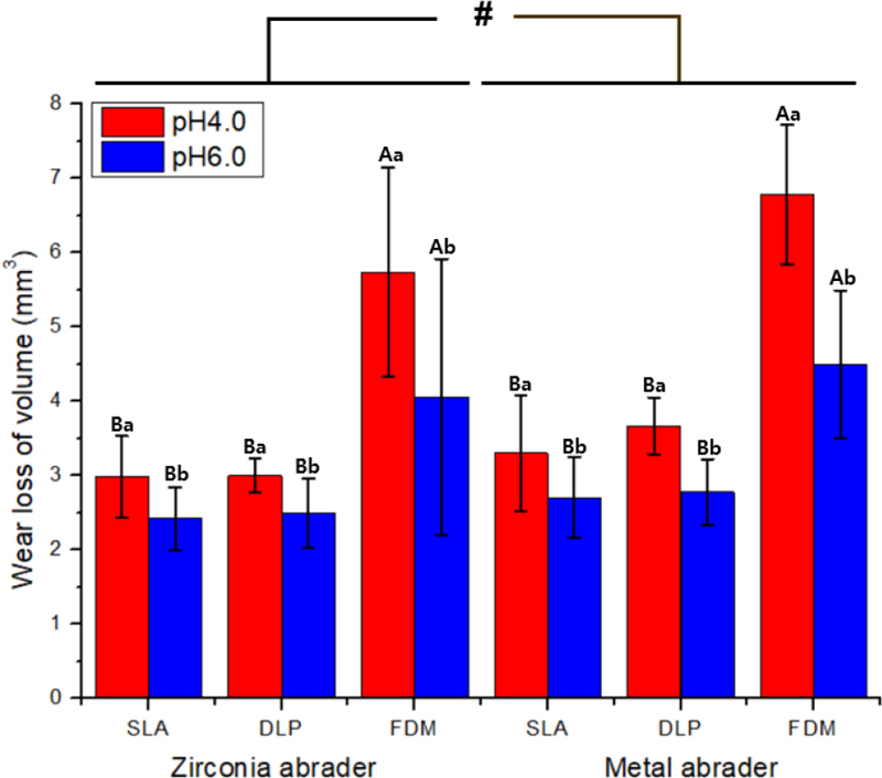
The wear loss of volume of the 3D-printed specimens after the chewing simulations is presented in Table 2 and Figure 1. The mean±standard deviations values for wear loss of volume against the zirconia abrader in the presence of pH 6.0 was 2.42±0.42 mm3 for the SLA group, 2.49±0.47 mm3 for the DLP group and 4.06±1.86 mm3 for the FDM group. For pH 4.0, the results were 2.98±0.55 mm3 for the SLA group, 3.00±0.23 mm3 for the DLP group and 5.74±1.41 mm3 for the FDM group. There were no significant differences in the volume loss between the SLA and DLP group in both pH (p>0.05). However, the FDM group showed significant differences with both the SLA and DLP group in both pH (p<0.05). In addition, all specimens immersed in artificial saliva of pH 4.0 resulted in significantly more wear loss of volume than specimens immersed in pH 6.0 (p<0.05), regardless of AM technology.

Wear loss of volume expressed as mean±standard deviations for specimens in the presence of different pH values. The same capital letters indicate no statistically significant difference between results in accordance with additive manufacturing (AM) methods (p<0.05). The same lower-case letters indicate no statistically significant difference between specimens immersed in pH 4.0 and pH 6.0 (p<0.05).

Wear loss of volume for specimens in the presence of different pH values, with different abrader (zirconia and metal). # indicates statically significant greater wear loss of volume compared to other type of abrader (p<0.05).
The mean±standard deviations values for wear loss of volume against the metal abrader in the presence of pH 6.0 was 2.70±0.54 mm3 for the SLA group, 2.78±0.44 mm3 for the DLP group and 4.09±0.99 mm3 for the FDM group. In the presence of pH 4.0, the results were 3.30±0.78 mm3 for the SLA group, 3.67±0.38 mm3 for the DLP group and 6.78±0.94 mm3 for the FDM group. Similar to the results when zirconia abrader was used, there were no significant differences in the volume loss between the SLA and DLP group (p>0.05) in both pH. However, the FDM group showed significant differences with both the SLA and DLP group (p<0.05) in both pH. In addition, all specimens immersed in artificial saliva of pH 4.0 resulted in significantly more wear loss of volume than specimens immersed in pH 6.0 (p<0.05), regardless of AM technology.
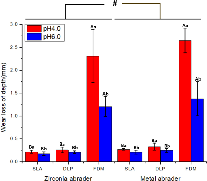
The wear loss of maximal depth of the 3D-printed specimens after the chewing simulations is presented in Table 3 and Figure 2. The mean wear loss of maximal depth±standard deviations against the zirconia abrader in the presence of pH 6.0 was 0.18±0.04 mm for the SLA group, 0.21±0.03 mm for the DLP group and 1.21±0.22 mm for the FDM group. In the presence of pH 4.0, the results were 0.22±0.03 mm for the SLA group, 0.26±0.06 mm for the DLP group and 2.31±0.58 mm for the FDM group. There were no significant differences in the wear loss of maximal depth between the SLA and DLP group for both pH (p>0.05). However, the FDM group showed significant differences with both the SLA and DLP group for both pH (p<0.05). In addition, all specimens immersed in artificial saliva of pH 4.0 resulted in significantly more wear loss of maximal depth than specimens immersed in pH 6.0 (p<0.05), regardless of AM technology.

Wear loss of maximal depth expressed as mean±standard deviations for specimens in the presence of different pH values. The same capital letters indicate no statistically significant difference between results in accordance with additive manufacturing (AM) methods. The same lower-case letters indicate no statistically significant difference between specimens immersed in pH 4.0 and pH 6.0 (p<0.05).

Wear loss of maximal depth for specimens in the presence of different pH values, with different abrader (zirconia and metal). # indicates statically significant greater wear loss of volume compared to other type of abrader (p<0.05).
The mean wear loss of maximal depth±standard deviations against the metal abrader in the presence of pH 6.0 was 0.21±0.04 mm for the SLA group, 0.25±0.04 mm for the DLP group and 1.38±0.37 mm for the FDM group. In the presence of pH 4.0, the results were 0.27±0.02 mm for the SLA group, 0.33±0.08 mm for the DLP group and 2.65±0.27 mm for the FDM group. There were no significant differences in the wear loss of maximal depth between the SLA and DLP group in both pH (p>0.05). However, the FDM group showed significant differences with both the SLA and DLP group, in both pH (p<0.05). In addition, all specimens immersed in artificial saliva of pH 4.0 resulted in significantly more wear loss of maximal depth than specimens immersed in pH 6.0 (p<0.05), regardless of AM technology.
2. Qualitative results of wear on the printed specimens
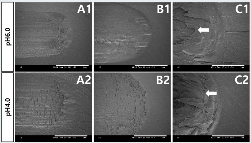
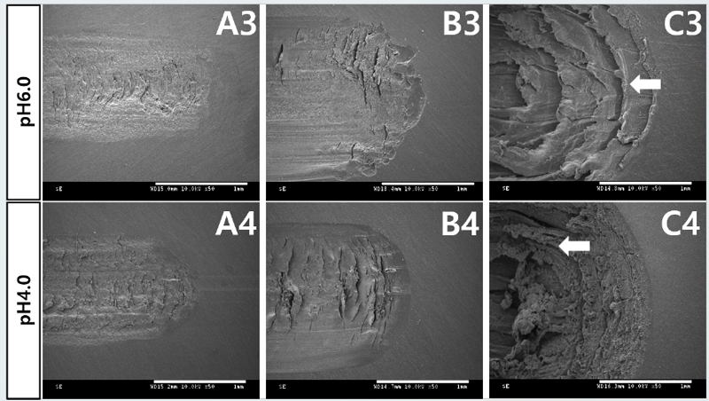
The FE-SEM images of the worn surfaces of the specimens after the wear tests are shown in Figure 3 and 4. All types of resin showed cracks and dented features. For the SLA resin specimens, a smooth surface with small cracks and grooves oriented parallel with the siding direction were observed [Figure 3 (A1) to (A4), and Figure 4 (A1) to (A4)]. The DLP resin specimens showed slightly wide range of grooves and dented fractures [Figure 3 (B1) to (B4) and Figure 4 (B1) to (B4)]. For the FDM resin specimens, remarkably showed more wide range of cracks and sharp step of the layer [white arrow in Figure 3 (C1) and (C2) and Figure 4 (C3) and (C4)]. In the presence of pH 6.0 specimens, a smooth surface with small steps on the fractured surface were seen for specimen [Figure 3 (A1), (B1) and (C1) and Figure 4 (A3), (B3) and (C3)], while pH 4.0 specimens remarkably distributed wrinkles-like appearance and patchy surface was observed [Figure 3 (A2), (B2) and (C2) and Figure 4 (A4), (B4) and (C4)]. Additionally, the surfaces of the wear areas of the three materials in contact with the zirconia abrader appeared to be relatively smoother than those in contact with the CoCr alloy abrader (Figure 3). For CoCr alloy abrader, the features relatively showed rough surface with faint lamellae (Figure 4). All resin specimens changed from smooth to rough surface with layering fracture. Based on FE-SEM findings there were variations in surface in term of different AM technologies type and different pH solutions influence, especially the appearance of surface layering fracture with acidic pH solutions and when used to CoCr alloy abrader.

FE-SEM images of the worn surfaces of the materials against the zirconia abrader. SLA specimens (A1-A2); DLP specimens (B1–B2); FDM specimens(C1–C2); under different pH solutions (4.0, and 6.0). White arrow indicates more wide range of cracks and sharp step of the layer. Scale bar is 1 mm.
Discussion
This study results demonstrated (1) influence in acidic pH and (2) difference in wear resistance among temporary resin specimens fabricated from different AM technologies, and, therefore, the null hypothesis was all rejected. In this study, the wear resistance were measured by varying the concentration of the pH solutions. All specimens tested in this study showed that acidic solutions of pH 4.0 decrease wear resistance which is in agreement with previous studies(16). Influence of acidic environment also showed worn surface after two body wear tests in the SEM images. In the presence of pH 6.0 specimens, a smooth surface with small steps on the fractured surface were seen for specimen [Figure 3 (A1), (B1) and (C1) and Figure 4 (A3), (B3), and (C3)], while pH 4.0 specimens remarkably distributed wrinkles-like appearance and patchy surface was observed [Figure 3 (A2, B2, C2), Figure 4 (A4, B4, C4)]. In addition, the results that there were no significant differences in the wear resistance between the SLA and DLP group, while FDM group had significantly lower values after immersion in acidic solution of pH 4.0. The reason that the PLA component printed by FDM have attributed this to the quick diffusion of acidic solution of pH 4.0 from interior of the high-porosity devices (9). When resins are absorbed in water, PLA had inferior moisture barrier compared with synthetic polymers (17), and easily hydrolyzed by moisture (18). So, management of moisture environment and hydrolytic degradation of PLA is significantly sensitive factors.
The wear resistance test was evaluated based on two body wear tests. The amount of wear of the SLA group was similar to the wear amount of the DLP group. The values of volume loss and maximal depth loss of wear showed similar patterns to each other. Just worn surface of the DLP group showed as slightly wide range of grooves and dented features in the SEM images [Figure 3 (B1, B2), Figure 4 (B3, B4)]. In the case of the FDM group, the maximal depth loss and volume loss values were remarkably large. Also, in SEM images remarkably showed more wide range of cracks and sharp step of the layer [white arrow in Figure 3 (C1, C2), Figure 4 (C3, C4)]. Because the PLA components printed by FDM printer tend to be weak and result in lower impact resistance (19). The most influential manufacturing factors affecting the wear performance of the FDM parts are raster angle, layer thickness, build orientation and the number of contours. Differences in wear patterns were found between the materials depending on the abraders. When looking at the specimens, volume loss and depth loss deviation were generally lower when zirconia was used than when the CoCr alloy was used as the abraders. Additionally, the surfaces of the wear areas of the specimens in contact with the zirconia abrader appeared relatively smooth in the SEM images. The differences in the results may be because of the presence or absence of the fillers and the nature of the fillers (20). Also the CoCr alloy used in DMLS method was during solidification of melting metal in particular “Co” can enhance crack tendency (21). The two body wear test parameters were as similar to the clinical loading conditions as possible. The load of 5 kg was equivalent to the average masticatory force of 49 N each. In addition, 0.4 to 0.75 N ranges of force and 10,000 to 1,200,000 cycles are adopted for most wear tests. A cycle of 250,000 loadings is similar to about one year in clinical situations (22). Thus, 20,000 cycles of load are comparable with approximately one of chewing, from a clinical perspective. However, depending on the condition of the patient, the temporary restorations may be multiple units of prosthesis, and depending on periodontal or implant surgery, the period of use may be extended.
This study had a limitation in that it is an in vitro study. These specimens had a flat surface, whereas teeth and restorations have complicated shapes that cause different stresses at various sites on the restoration surface (23). Another limitation is wear resistance test that three body wear test may be even more clinically important compared to two body wear tests (24). Such three body wear tests may give meaningful results in terms of prediction of clinical performance, however, are likely to be of limited help in terms of materials characteristic.
In our study, the wear resistance of the materials manufactured using three different types of 3D printed resin was evaluated, and the results showed that the SLA and DLP specimens could yield stable clinical outcomes in comparison to those of the FDM specimens. The FDM printer was easy to handle in the clinic but PLA investigations over the last decade have revealed that standard 3D-printed PLA has poor mechanical qualities (19). Further studies are needed to evaluate FDM resin using objective data. In additions, in the presence of acidic pH demonstrated in poor wear resistance. Thus, management of acidic in oral environment is significantly sensitive factors to be considered.
Conclusions
The aim of this study was to influence of acidic pH and wear resistance of fabricated temporary based on resin specimens through different AM technologies. With the limitation that this was an in vitro experiment, the results showed that the SLA and DLP group could yield stable clinical outcomes compared to those of the FDM group. In addition, in the presence of acidic pH showed in poor wear resistance results. Thus, acidic in oral environments significantly to be needed for attention. For a wider application of the 3D printing technology in dentistry, additional studies are required to examine flexural strength, surface hardness. These mechanical properties of the 3D printed resin materials with respect to many factors should be studied in the future.
Acknowledgments
This work was supported by the National Research Foundation of Korea (NRF) grant funded by the Korean government (MSIT) (No. 2021R1A2C2091260).
References
-
Katreva I, Dikova T, Abadzhiev M, Tonchev T, Dzhendov D, Simov M, et al. 3D-printing in contemporary prosthodontic treatment. Scripta Scientifica Medicinae Dentalis. 2016;2(1):7-11.
[https://doi.org/10.14748/ssmd.v1i1.1446]

-
Schweiger J, Edelhoff D, Güth J-F. 3D Printing in Digital Prosthetic Dentistry: An Overview of Recent Developments in Additive Manufacturing. J Clin Med. 2021;10(9):2010.
[https://doi.org/10.3390/jcm10092010]

-
Tian Y, Chen C, Xu X, Wang J, Hou X, Li K, et al. A review of 3D printing in dentistry: Technologies, affecting factors, and applications. Scanning. 2021;2021.
[https://doi.org/10.1155/2021/9950131]

-
Cha HS, Park JM, Kim TH, Lee JH. Wear resistance of 3D-printed denture tooth resin opposing zirconia and metal antagonists. J Prosthet Dent. 2020;124(3):387-94.
[https://doi.org/10.1016/j.prosdent.2019.09.004]

-
Aldahian N, Khan R, Mustafa M, Vohra F, Alrahlah A. Influence of Conventional, CAD-CAM, and 3D Printing Fabrication Techniques on the Marginal Integrity and Surface Roughness and Wear of Interim Crowns. Appl Sci (Basel). 2021;11(19):8964.
[https://doi.org/10.3390/app11198964]

-
Abdullah AO, Pollington S, Liu Y. Comparison between direct chairside and digitally fabricated temporary crowns. Dent Mater J. 2018;37(6):957-63.
[https://doi.org/10.4012/dmj.2017-315]

-
Firlej M, Pieniak D, Niewczas AM, Walczak A, Domagała I, Borucka A, et al. Effect of artificial aging on mechanical and tribological properties of CAD/CAM composite materials used in dentistry. Materials (Basel). 2021;14(16):4678.
[https://doi.org/10.3390/ma14164678]

-
Cilli R, Pereira JC, Prakki A. Properties of dental resins submitted to pH catalysed hydrolysis. J Dent. 2012;40(12):1144-50.
[https://doi.org/10.1016/j.jdent.2012.09.012]

-
Farah S, Anderson DG, Langer R. Physical and mechanical properties of PLA, and their functions in widespread applications—A comprehensive review. Adv Drug Deliv Rev. 2016;107:367-92.
[https://doi.org/10.1016/j.addr.2016.06.012]

-
Drummond JL, Lin L, Al-Turki LA, Hurley RK. Fatigue behaviour of dental composite materials. J Dent. 2009;37(5):321-30.
[https://doi.org/10.1016/j.jdent.2008.12.008]

-
Rahim TNAT, Mohamad D, Akil HM, Ab Rahman I. Water sorption characteristics of restorative dental composites immersed in acidic drinks. Dent Mater. 2012;28(6):e63-e70.
[https://doi.org/10.1016/j.dental.2012.03.011]

-
Park J-M, Ahn J-S, Cha H-S, Lee J-H. Wear resistance of 3D printing resin material opposing zirconia and metal antagonists. Materials (Basel). 2018;11(6):1043.
[https://doi.org/10.3390/ma11061043]

-
Myagmar G, Lee J-H, Ahn J-S, Yeo I-SL, Yoon H-I, Han J-S. Wear of 3D printed and CAD/CAM milled interim resin materials after chewing simulation. J Adv Prosthodont. 2021;13(3):144.
[https://doi.org/10.4047/jap.2021.13.3.144]

-
Kim M, Lee M, Kim K, Yang S, Seo J, Choi S, et al. Enamel Demineralization Resistance and Remineralization by Various Fluoride-Releasing Dental Restorative Materials. Materials (Basel). 2021;14(16).
[https://doi.org/10.3390/ma14164554]

-
Krejci I, Lutz F, Reimer M, Heinzmann J. Wear of ceramic inlays, their enamel antagonists, and luting cements. J Prosthet Dent. 1993;69(4):425-30.
[https://doi.org/10.1016/0022-3913(93)90192-Q]

-
Yilmaz EÇ, Sadeler R, Duymuş ZY, Özdoğan A. Effect of ambient pH and different chewing cycle of contact wear on dental composite material. Dentistry and Medical Research. 2018;6(2):46-50.
[https://doi.org/10.4103/dmr.dmr_26_18]

-
Pan P, Liang Z, Zhu B, Dong T, Inoue Y. Roles of physical aging on crystallization kinetics and induction period of poly (L-lactide). Macromolecules. 2008;41(21):8011-9.
[https://doi.org/10.1021/ma801436f]

-
Yew G, Yusof AM, Ishak ZM, Ishiaku U. Water absorption and enzymatic degradation of poly (lactic acid)/rice starch composites. Polym Degrad Stab. 2005;90(3):488-500.
[https://doi.org/10.1016/j.polymdegradstab.2005.04.006]

-
Subramaniyan M, Karuppan S, Radhakrishnan K, Kumar RR, Kumar KS. Investigation of wear properties of 3D-printed PLA components using sandwich structure–A review. Materials Today: Proceedings. 2022.
[https://doi.org/10.1016/j.matpr.2022.04.913]

-
Cha H-S, Park J-M, Kim T-H, Lee J-H. Wear resistance of 3D-printed denture tooth resin opposing zirconia and metal antagonists. J Prosthet Dent. 2020;124(3):387-94.
[https://doi.org/10.1016/j.prosdent.2019.09.004]

-
Béreš M, Silva C, Sarvezuk P, Wu L, Antunes L, Jardini A, et al. Mechanical and phase transformation behaviour of biomedical Co-Cr-Mo alloy fabricated by direct metal laser sintering. Materials Science and Engineering: A. 2018;714:36-42.
[https://doi.org/10.1016/j.msea.2017.12.087]

-
DeLong R, Sakaguchi R, Douglas WH, Pintado M. The wear of dental amalgam in an artificial mouth: a clinical correlation. Dent Mater. 1985;1(6):238-42.
[https://doi.org/10.1016/S0109-5641(85)80050-6]

-
Mair L, Stolarski T, Vowles R, Lloyd C. Wear: mechanisms, manifestations and measurement. Report of a workshop. J Dent. 1996;24(1-2):141-8.
[https://doi.org/10.1016/0300-5712(95)00043-7]

-
McCabe J, Molyvda S, Rolland S, Rusby S, Carrick T. Two-and three-body wear of dental restorative materials. Int Dent J. 2002;52:406-16.
[https://doi.org/10.1111/j.1875-595X.2002.tb00730.x]


