Influence of depth of cavity on color blending effect of structurally colored resin
Abstract
Omnichroma (OMN) is a recently introduced structurally colored resin composite that expresses color based on the tooth structure surrounding the cavity. This study aims to investigate the effects of varying the cavity depth on the color blending of OMN.
Conventional resin composite (Filtek Z250 in the A2, A3, and A4 shades) and structurally colored resin (Omnichroma) were used. Two types of specimens were prepared using custom silicone molds (diameter: 8 mm, thickness: 2, 3, 4 mm). Single specimens (diameter: 8 mm) comprised only Z250 in the A2, A3, A4 or OMN (n=10 each). Dual specimens comprised an outer ring (diameter : 8 mm) of Z250 in the A2, A3, or A4 and an inner hole (diameter: 4 mm) filled with OMN to different depths (1, 2, 3, or 4 mm, n=10 per shade per thickness). The colors were measured using the Commission Internationale d’Eclairage (CIE) L*a*b* system. Color differences (ΔE) according to the cavity depth and translucency parameter were measured.
The ΔE values of dual specimens with the A2, A3, and A4 shades of Z250 were 1.66–5.07, 0.50–2.57, and 1.26–3.48, respectively. At the same specimen thickness, ΔE increased with increasing cavity depth. At the same cavity depth, ΔE increased with decreasing thickness of the bottom of the restoration. The highest translucency parameter was observed for 2 mm-thick OMN. The color blending of OMN increased with decreasing cavity depth in specimens of the same thickness, and with increasing bottom thickness of the restoration at the same cavity depth.
초록
Omnichroma (OMN)는 최근에 소개된 구조색으로 색상을 나타내는 복합레진으로 와동을 둘러싼 치아 구조를 기반으로 색상을 표현한다. 본 연구의 목적은 다양한 와동의 깊이가 OMN의 색상 혼합 효과에 미치는 영향을 조사하는 것이다. 기존의 복합 레진(Filtek Z250 A2, A3, A4 색상)과 구조색 기반 복합레진(Omnichroma)을 사용하였다. 자체 제작한 실리콘 몰드(직경: 8 mm, 두께: 2, 3, 4 mm)를 사용하여 두 가지 유형의 시편을 준비하였다. 단일 시편(직경: 8 mm)은 Z250의 A2, A3, A4 색상 또는 OMN으로만 충전하여 각각 10개씩 만들었다. 이중 시편은 Z250의 A2, A3, A4 색상으로 직경 8mm의 디스크 형태의 시편을 만든 후 직경이 4 mm인 내부 와동을 1, 2, 3, 4 mm 깊이로 형성하여 OMN을 충전하였다. 이중 시편은 내부 와동의 두께당, 디스크 색상의 종류당 각각 10개씩의 시편을 제작하였다. 색상은 CIE (Commission Internationale d'Eclairage) L*a*b* 시스템을 사용하여 와동 깊이와 반투명도에 따른 색 변화 값(ΔE)을 측정하였다. Z250의 A2, A3, A4 색상으로 충전한 이중 시편의 색 변화 값(ΔE)은 각각 1.66~5.07, 0.50~2.57, 1.26~3.48 였다. 동일한 시편 두께에서 와동의 깊이가 증가함에 따라 색 변화 값(ΔE)이 증가하였고 동일한 와동 깊이에서 수복물 하부 구조물의 두께가 감소함에 따라 색 변화 값(ΔE)은 증가하였다. 또한 2 mm 두께의 OMN에서 가장 높은 반투명도가 나타났다. 결론적으로, OMN의 색상 혼합 효과는 동일한 두께의 시편에서 와동 깊이가 감소함에 따라 증가하였고, 동일한 와동 깊이에서 수복물 하부 구조물의 두께가 증가함에 따라 증가하였다.
Keywords:
Structurally colored resin, Omnichroma, Blending effect, Cavity depth키워드:
구조색 복합 레진, 색상 혼합 효과, 와동 깊이Introduction
Since Bowen introduced resin composites to dentistry using Bis-GMA as a matrix in the 1960s, composite resin materials have been widely used in dental restorations. Owing to advances in their chemical and mechanical properties as well as their aesthetic advantages over other materials, resin materials have replaced traditionally used gold and amalgam (1) and are being increasingly used for posterior teeth restorations. Interestingly, though not a primary criterion, aesthetics—particularly color matching with adjacent teeth—have been found to be a higher priority for material selection than criteria such as durability, manipulability, polishability, and wear resistance (2).
The shade of natural teeth is determined by both opacity and color, which in turn is determined by the combination of hue, chroma, and value (3). However, in the shade matching of resin composites, optical properties such as translucency, fluorescence, and opalescence also have an impact, as well as its surface gloss and the reflection, refraction, transmission, absorption, and scattering of light (4). Consequently, even if commercial resin composites are labeled as the same color based on their chroma and hue, their actual color can vary according to the manufacturer and product. (5, 6). Although the Vita classical shade guide, wherein colors are classified and assigned names from A1 to D4, is used as the standard method for selecting resin shade (7), shade matching for posterior restorations is challenging for clinicians because it is difficult to match the shade in the dark oral cavities (2). Various layering techniques have also been used to match the shade and optical properties of resin composites to those of the opaque dentin, transparent dentin, enamel, and incisal edge of natural teeth based on various specific optical parameters (8). However, these layering techniques are subjective and sensitive, making them challenging for clinicians to use.
There are two mechanisms by which resin expresses color (9, 10). The first, chemical color, is the most common type of color expression in composite resins and involves the use of dyes and pigments (11); the color is determined by the wavelength of light transmitted, reflected, or absorbed by the pigment. The second is structural color, which is based on the interaction of incident light with nano-structured fillers; the coloring results from optical processes, such as diffraction, interference, and scattering (12, 13). Therefore, structural color does not fade easily, unlike chemical color.
There are two methods for measuring color: the method of visual observation and the method using a color measurement device. A spectrophotometer is a device used to obtain objective information about color appearance by measuring the L*a*b* values of a specimen, allowing for the quantification of color. This study focuses on assessing the degree of blending with surrounding colors when restoring structurally colored resin based on the depth of cavity. Therefore, when using the formalized ΔE, which represents the objective numerical L*a*b* values of the specimen, the value can also be quantified, enabling an objective assessment of the blending effect.
The color blending effect, also known as the chameleon effect, refers to a phenomenon in which the color difference between the restorative material and the surrounding tooth structure is perceived to be smaller when they are physically adjacent to each other (14). Factors that affect the chameleon effect include the type and shade of the resin composite, degree of the color difference between the tooth and the restoration, and size and thickness of the restoration (15, 16). After restoring a cavity with a resin composite, the color and optical properties of the resin composite, as well as the bottom structure of the restoration, all affect the interaction of light. This results in the chameleon effect of the restoration (17). Translucency, defined as the relative amount of light that passes through an object after being absorbed and scattered (18), is also an important factor when evaluating the color blending effect (13). Therefore, when the translucence parameter (TP) is low, structurally colored resins, which express color by reflecting, absorbing, and scattering light reflected from the structure surrounding the cavity, are affected less by the color of the surrounding structure.
Omnichroma (OMN), a recently introduced single-shade resin, is the first commercially available structurally colored resin. OMN is composed of uniform supra-nano spherical fillers of SiO2 and ZrO2 with a particle size of 260 nm (smaller than the wavelength of visible light, ~380 nm) (12, 19). Unlike resin composites that contain pigments, OMN is a structurally colored resin composite that exhibits the chameleon effect by expressing color based on the structure surrounding the cavity (10). The fillers change from red to a yellow color when light passes through, and, combined with the reflected color of the structure surrounding the restoration, present the ideal shade for dental restoration (14), thereby saving shade-matching time for clinicians and decreasing chair time for patients. Furthermore, the use of OMN allows for shade matching to be conducted with only one single-shade resin composite material, which reduces the time and effort required for shade matching. Therefore, OMN is increasingly gaining attention as an alternative to chemically colored resin composites.
Structurally colored resin can express different colors depending on the amount of structure surrounding the cavity, specifically the depth of the cavity. (10) However, despite studies having been conducted on the effects of cavity depth and background colors on the color blending of OMN (17, 19, 20), there have been no reports on the relationship between the color blending of OMN and the cavity depth and amount of surrounding structure. Therefore, in this study, we investigated the effects of varying the cavity depth and, consequently, the amount of surrounding structure on the color blending of OMN. The null hypothesis was that the difference in cavity depth does not affect the color blending of OMN.
Materials and Methods
1. Preparation of samples
OMN, a structurally colored resin composite, and the A2, A3, and A4 shades of Filtek Z250, a pigment-colored resin composite, were used. Table 1 lists the manufacturer, composition, and shade of each product. Single specimens with only one resin and dual specimens with both resins were made using a customized disk-shaped mold. For the single specimens, disk-shaped specimens with a diameter of 8 mm and thicknesses of 2, 3, and 4 mm were prepared with each type and shade of resin (n=10 each). For the dual specimens, the outer rings (diameter 8 mm, thickness 2, 3, and 4 mm) were filled with Z250 in the A2, A3, or A4 shade, and the inner hole (diameter 4 mm) was filled with different thicknesses (1, 2, 3, and 4 mm) of OMN (n=10 per shade per thickness) (Figure 1). When filling the resin into the mold, Z250 was filled in 2-mm increments and OMN was filled in 3-mm increments according to the manufacturer's instructions. After the mold was filled with resin, a glass slide was placed on top of it to flatten it, and light-curing was performed for 20 s using an LED light unit (Bluephase Style 20i, Ivoclar Vivadent, Lichenstein, Germany) after each increment. Subsequently, the top surfaces of the specimens were polished with a grinder (Metaserve 250 grinder-polisher; Buehler, IL, USA) and 400-, 600-, and 1,200-grit sandpaper. The thicknesses of the specimens were measured with a digital caliper.
2. Measurement of color
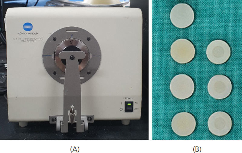
The center of the top surface of each specimen was measured for the Commission Internationale d’Eclairage (CIE) L*a*b* parameters using a spectrophotometer (CM-3600d, Minolta, Tokyo, Japan) on white and black ceramic backgrounds (Figure 2). Before measuring each sample, the spectrophotometer was calibrated with the standard calibrating blocks (white and black) according to the manufacturer's instructions.
3. Calculation of the color difference and translucency parameter
The color difference value, ΔE, is the difference between the colors of the single and dual specimens with different OMN thicknesses and the same overall thickness and outer-ring shade. ΔE was calculated with the L*a*b* values measured from all the specimens using the following formula:
The translucency parameter (TP) was calculated with the L*a*b* values measured on black and white backgrounds for the OMN, A2, A3, and A4 single specimens with thicknesses of 2, 3, and 4 mm using the following formula:
“B” and “W” represent black and white, respectively.
4. Statistical analysis
The data were obtained using SPSS 29.0 (SPSS Inc. Chicago, IL, USA). The color differences were analyzed by one-way analysis of variance, Scheffe's post hoc test, and student’s t-test. The significance level was set to 0.05.
Result
1. Color difference (ΔE)
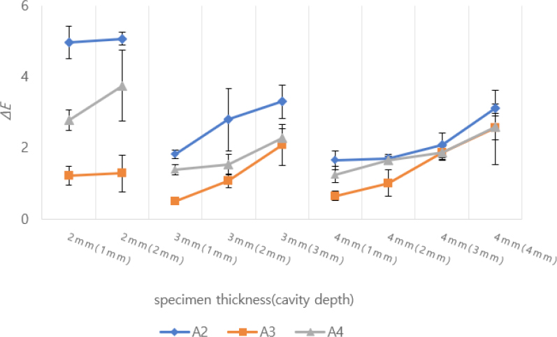
Table 2 lists the L*, a*, and b* values according to shade, specimen thickness, and cavity depth. Figure 3 shows the ΔE values of all specimens according to specimen thickness and cavity depth. The ΔE values for specimens A2, A3, and A4 were 1.66–5.07, 0.50–2.57, and 1.26–3.48, respectively.
For the A2 specimens, the ΔE values increased significantly with increasing cavity depth at the same specimen thickness, except when the cavity depth increased from 2 to 3 mm for the 3 mm-thick specimens and from 1 to 2 mm for the 4 mm-thick specimens. Meanwhile, the ΔE values decreased significantly with increasing specimen thickness at the same cavity depth, except when the specimen thickness increased from 3 to 4 mm at a 1-mm cavity depth (Table 3).
For the A3 specimens, the ΔE values increased significantly with increasing cavity depth in all specimens of the same thickness, except when the cavity depth increased from 1 to 2 mm for the 4 mm-thick specimens. Meanwhile, for the same cavity depth, the ΔE values decreased significantly only for an increase in specimen thickness from 2 to 3 mm at a 1-mm cavity depth and an increase from 3 to 4 mm at a 3-mm cavity depth (Table 4).
For the A4 specimens, the ΔE values significantly increased only for the 2 mm-thick specimens when the cavity depth increased from 1 to 2 mm and for the 3 mm-thick specimens when the cavity depth increased from 2 to 3 mm. Furthermore, the ΔE values decreased significantly with increasing specimen thickness at the same cavity depth, except when the specimen thickness was increased from 3 to 4 mm at cavity depths of 1 mm and 2 mm (Table 5).
2. Translucency parameter (TP)
Table 6 lists the TP values for the OMN, A2, A3, and A4 single specimens with 2-, 3-, and 4-mm thicknesses. In all specimens, the TP decreased significantly with increasing specimen thickness. The OMN specimens had higher TP values than the A2, A3, and A4 specimens at all thicknesses. The A3 specimens had a lower TP than the A2 specimens at all specimen thicknesses except 3 mm but did not have a higher TP than the A4 specimens at any thickness.
Discussion
Previous studies reported that the blending effect varies according to the resin composite product and shade and increases as the restoration size decreases and transparency increases; (5, 6, 16) restoration size refers to both the width and thickness of the restoration. Many studies have focused on the blending effect of structurally colored resin according to differences in the restoration width (10, 21, 22). However, the restoration color is affected by the surface color as well as the light scattering and reflectivity of the background of the deep cavity. The thickness of the restoration can also have a significant effect (23). Therefore, in this study, the restoration thickness was adjusted by varying the cavity depth, and the ΔE values were measured.
Among the dual specimens, the specimen with the A3 shade showed the lowest ΔE value of OMN. This was attributed to the similarity between the inherent shades of OMN and A3 Z250. Paravina et al. reported that a smaller initial color difference between the resin composite and the surrounding structure of the cavity indicates a greater blending effect (15). Therefore, the color blending effect of OMN with the A3 shade was superior to that with the A2 and A4 shades. In addition, the bottom thicknesses of the dual specimens with A3 shade did not have a significant effect on the ΔE values because of the excellent blending effect of OMN with the A3 shade of Z250. However, the dual specimens with A2 and A4 shade showed higher ΔE values at a specimen thickness of 2 mm than at 3 mm and 4 mm, which may be due to an insufficient amount of structure surrounding the cavity to mimic the color from.
In most of the specimens, at the same specimen thickness, a significant increase in ΔE values was observed when the cavity depth increased by 2 or 3 mm regardless of the shade, which did not occur when the cavity depth increased by 1 mm. This indicates that cavity depth is an important factor influencing the color blending of OMN.
At a cavity depth of 1 mm, all dual specimens showed a significant decrease in ΔE values when the specimen thickness increased by 2 mm. However, when the cavity depth was 2 mm, the ΔE values decreased significantly when the specimen thicknesses were increased by 2 mm, except for the A3 dual specimen. These results can be explained as follows: when the overall specimen thickness increased by 2 mm rather than 1 mm at the same cavity depth, there was a greater increase in the bottom thickness of the cavity structure, which resulted in a significant increase in the ΔE values. Therefore, it can be concluded that the bottom thickness of the cavity structure is an important factor influencing the color blending of OMN.
The ΔE values of OMN varied depending on the shade of Z250 in the dual specimens. These results are consistent with those reported by Pereira Sanchez et al., who concluded that the blending effect varies according to shade and decreases as the size of the restoration increases (6). Increasing the restoration thickness by adjusting the cavity depth resulted in an increase in the ΔE value of OMN, indicating decreased blending effect. That is, OMN showed an increase in the blending effect as the restoration size decreased. The null hypothesis was therefore rejected, as varying the cavity depth affected the color blending of OMN. Similar results are expected when OMN is clinically applied as teeth restorations.
OMN had a higher TP than the A2, A3, and A4 shades of Z250 at all thicknesses. Hence, OMN could reflect, absorb, and scatter light such that the color of the structure surrounding the cavity was better matched than when using Z250 of the A2, A3, or A4 shade. The TP of OMN decreased with increasing specimen thickness, leading to a decrease in its ability to reflect, absorb, and scatter the color of the structure surrounding the cavity; this decreased its ability to mimic the color of the surrounding structure. Therefore, as the thickness of OMN increased, the ΔE value increased whereas the blending effect decreased.
This is a rare study that determined the effect of cavity depth on the shade of structurally colored resin composites, as opposed to that of cavity width, as has been reported thus far. This study also showed that the color difference decreased with decreasing cavity size, which is in agreement with previous studies on the blending effect (15, 16, 17). In addition, at the same specimen thickness, the blending effect increased with decreasing cavity depth.
According to Inokoshi et al., the ΔE limit under which the color difference is undetectable by the human eye is ΔE < 3.3 (24). As the ΔE value of OMN for most specimens was ΔE < 3.3, we can conclude that the structurally colored resin OMN shows potential as an excellent alternative to conventional chemically colored resin composites, which require complex shade-matching. The limitation of this study is that only one structurally colored resin was used, and additional evaluation on the color blending effect of other structurally colored resins is needed. Additionally, further study on the long-term color stability of OMN is required to accurately judge its practical applicability as dental restorations.
Conclusion
In this study, the structurally colored resin composite, OMN, exhibited an increased blending effect when the cavity depth was decreased at the same specimen thickness. Furthermore, specimens with a greater bottom thickness exhibited an increased blending effect at the same cavity depth.
Acknowledgments
This work was supported by a 2-year Research Grant of Pusan National University
References
-
Ferracane JL. Resin composite--state of the art. Dent Mater. 2011;27(1):29-38.
[https://doi.org/10.1016/j.dental.2010.10.020]

- Hervás-García A, Martínez-Lozano MA, Cabanes-Vila J, Barjau-Escribano A, Fos-Galve P. Composite resins. A review of the materials and clinical indications. Med. Oral Patol. Oral Cir. Bucal. 2006;11(2):E215–220
-
Jouhar R, Ahmed MA, Khurshid Z. An overview of shade selection in clinical dentistry. Appl Sci. 2022;12(14):6841.
[https://doi.org/10.3390/app12146841]

-
Lee YK. Influence of scattering/absorption characteristics on the color of resin composites. Dent Mater. 2007;23(1):124-31.
[https://doi.org/10.1016/j.dental.2006.01.007]

-
Lee YK, Yu B, Lee SH, Cho MS, Lee CY, Lim HN. Shade compatibility of esthetic restorative materials—A review. Dent Mater. 2010;26(12):1119-26.
[https://doi.org/10.1016/j.dental.2010.08.004]

-
Pereira Sanchez N, Powers JM, Paravina RD. Instrumental and visual evaluation of the color adjustment potential of resin composites. J Esthet Restor Dent. 2019;31(5):465-70.
[https://doi.org/10.1111/jerd.12488]

-
Da Costa J, Fox P, Ferracane J. Comparison of various resin composite shades and layering technique with a shade guide. J Esthet Restor Dent. 2010;22(2):114-24.
[https://doi.org/10.1111/j.1708-8240.2010.00322.x]

-
Dietschi D, Fahl N. Shading concepts and layering techniques to master direct anterior composite restorations: an update. Br Dent J. 2016;221(12):765-71.
[https://doi.org/10.1038/sj.bdj.2016.944]

-
Shang L, Gu Z, Zhao Y. Structural color materials in evolution. Mater Today. 2016;19(8):420-1.
[https://doi.org/10.1016/j.mattod.2016.03.004]

-
Makoto S, Kurokawa H, Takahashi N, Toshiki T, Ishii R, Shiratsuchi K, et al. Evaluation of color-matching ability of a structural colored resin composite. Oper Dent. 2021;46(3):306–15.
[https://doi.org/10.2341/20-002-L]

-
Brainard DH, Cottaris NP, Radonjić A. The perception of colour and material in naturalistic tasks. Interface Focus. 2018;8(4):20180012-2.
[https://doi.org/10.1098/rsfs.2018.0012]

-
Yamaguchi S, Karaer O, Lee C, Sakai T, Imazato S. Color matching ability of resin composites incorporating supra-nano spherical filler producing structural color. Dent Mater. 2021;37(5):e269-75.
[https://doi.org/10.1016/j.dental.2021.01.023]

-
Vattanaseangsiri T, Khawpongampai A, Sittipholvanichkul P, Jittapiromsak N, Posritong S, Wayakanon K. Influence of restorative material translucency on the chameleon effect. Sci Rep-UK. 2022;12(1):8871.
[https://doi.org/10.1038/s41598-022-12983-y]

- Sharma N, Praveen S, Samant. Omnichroma: the see-it-to-believe-it technology. EAS J. Dent. Oral Med. 2021;3(3):100-4.
-
Paravina RD, Westland S, Kimura M, Powers JM, Imai FH. Color interaction of dental materials: Blending effect of layered composites. Dent Mater. 2006;22(10):903–8.
[https://doi.org/10.1016/j.dental.2005.11.018]

-
Paravina RD, Westland S, Imai FH, Kimura M, Powers JM. Evaluation of blending effect of composites related to restoration size. Dent Mater.2006;22(4):299-307.
[https://doi.org/10.1016/j.dental.2005.04.022]

-
Islam MS, Huda N, Mahendran S, Aryal AC S, Nassar M, Rahman MM. The blending effect of single-shade composite with different shades of conventional resin composites—An in vitro study. Eur J Dent. (2022)
[https://doi.org/10.1055/s-0042-1744369]

-
Gigilashvili D, Thomas JB, Hardeberg JY, Pedersen M. Translucency perception: A review. J Vision. 2021;21(8):4-4.
[https://doi.org/10.1167/jov.21.8.4]

-
Durand LB, Ruiz‐López J, Perez BG, Ionescu AM, Carrillo‐Pérez F, Ghinea R, et al. Color, lightness, chroma, hue, and translucency adjustment potential of resin composites using CIEDE2000 color difference formula. J Esthet Restor Dent. 2021;33(6):836-843.
[https://doi.org/10.1111/jerd.12689]

-
Abreu JLB, Sampaio CS, Benalcázar Jalkh EB, Hirata R. Analysis of the color matching of universal resin composites in anterior restorations. J Esthet Restor Dent. 2020;33(2):269-76.
[https://doi.org/10.1111/jerd.12659]

-
Ismail EH, Paravina RD. Color adjustment potential of resin composites: Optical illusion or physical reality, a comprehensive overview. J Esthet Restor Dent. 2022;34(1):42-54,
[https://doi.org/10.1111/jerd.12843]

-
Trifkovic B, Powers JM, Paravina RD. Color adjustment potential of resin composites. Clin oral invest. 2017;22(3):1601-7.
[https://doi.org/10.1007/s00784-017-2260-6]

-
Akgül S, Gündoğdu C, Bala O. Effects of storage time and restoration depth on instrumental color adjustment potential of universal resin composites. J Oral Sci. 2022;64(1): 49-52.
[https://doi.org/10.2334/josnusd.21-0290]

- S Inokoshi, Burrow MF, M Kataumi, Yamada T, Takatsu T. Opacity and color changes of tooth-colored restorative materials. Oper Dent. 1996;21(2):73-80.


