치약 성분이 미세 혼합형/나노필러 함유 복합레진의 칫솔질 내마모성에 미치는 영향
 ; Jung-Eun Park1
 ; Kyeong-Seon Kim2
 ; Tae-Hwan Kim1
 ; Chung-Cha Oh1
 ; Seung-O Ko33, *
 ; Min-Ho Lee1, *
초록
본 연구의 목적은 연마제 성분이 다른 치약들로 칫솔질 마모시험을 시행하여 미세 혼합형 및 나노필러 복합레진의 표면 형태와 표면 거칠기를 관찰하고, 내마모성을 비교하는 것이다. 미세 혼합형 필러가 함유된 2종의 복합레진(Z100 Restorative, Filtek Z250)과 나노필러가 함유된 1종의 복합레진 (Filtek Z350 XT)을 사용하였다. 칫솔질 마모시험을 위해 직경 10 mm, 두께 1 mm의 복합레진 시편 90개를 준비했다. pin-on-disk 구동방식의 마모시험기를 사용하여 작용력은 2 N, 100,000회 칫솔질을 수행하였다. 시험에 사용된 치약은 연마제 성분(하이드록시아파타이트, 탄산칼슘, 중탄산나트륨, 제올라이트-M)에 따라서 4그룹으로 분류하였다. 칫솔질 마모시험 후 시편의 표면 형태는 광학현미경과 주사전자현미경(SEM)으로 관찰하였고, 원자힘현미경(AFM)으로 표면 거칠기를 측정하였다. 미세 혼합형 복합레진 그룹들의 표면에서는 상대적으로 큰 마이크로 필러 입자들이 돌출되어 있고, 작은 분화구 형태의 결함들이 관찰되었다. 제올라이트-M을 함유한 치약으로 마모시험을 시행한 그룹들의 표면 거칠기 값이 유의하게 가장 높았다(P<0.05). 나노필러 함유 복합레진을 제올라이트-M이 함유된 치약으로 마모시험한 그룹에서 표면 거칠기 값이 유의하게(P<0.05) 가장 높았으나, 치약 종류와 상관없이 표면 거칠기가 0.1 µm 미만으로 낮은 값들을 나타냈다. 미세 혼합형 복합레진들의 표면에 비해 표면이 균일하고 매끄러워 보였다. 결과적으로, 나노필러 복합레진이 미세 혼합형 복합레진에 비해 상대적으로 더 높은 내마모성을 보였다. 이는 칫솔질 시의 마모 저항성이 재료의 임상적 내구성을 나타낼 수 있다.
Abstract
The purpose of this study was to observe the surface morphology and roughness of micro-hybrid and nano-filled resin composites and compare wear resistance by conducting a toothbrushing wear test with toothpastes with different abrasive ingredients. Two types of resin composites containing micro-hybrid fillers (Z100 Restorative, Filtek Z250) and one type of resin composite containing nanofillers (Filtek Z350 XT) were used. For the toothbrushing wear test, 90 resin composite samples with a diameter of 10 mm and a thickness of 1 mm were prepared. A force of 2 N and 100,000 cycles of brushing were performed using a pin-on-disk wear tester. The toothpastes used in the test were classified into 4 groups according to the abrasive ingredients (hydroxyapatite, calcium carbonate, sodium bicarbonate, and zeolite-M). After the toothbrushing wear test, the surface morphology of the samples was observed using an optical microscope and a scanning electron microscope (SEM), and the surface roughness was measured using atomic force microscopy (AFM). Relatively large filler particles (micro size) protruded from the surface of the micro-hybrid resin composite groups, and small crater-shaped defects were observed. The surface roughness values of the groups that performed the wear test with toothpaste containing zeolite-M were significantly higher than the other groups (P<0.05). The surface roughness value was significantly (P<0.05) highest in the group where the nanofilled resin composite was wear-tested with toothpaste containing zeolite-M. However, regardless of the type of toothpaste, the surface roughness showed low values of less than 0.1 μm. The surface appeared uniform and smooth compared to the surface of micro-hybrid resin composites. Finally, the nano-filled resin composite showed relatively higher wear resistance than the micro-hybrid resin composite. This means that wear resistance during brushing may indicate the durability of the material in the clinic.
Keywords:
Resin composite, Toothbrushing wear resistance, Surface roughness, Toothpaste키워드:
복합레진, 칫솔질 내마모성, 표면거칠기, 치약서론
치과 임상 분야에서 과거 금속재료가 제공해온 단순한 수복의 기능 뿐만아니라 심미성까지 만족시킬 수 있는 심미성 수복재료에 대한 요구가 증가했다(1). 최근 수복 치료에서는 뛰어난 심미성과 우수한 조작성을 지닌 복합레진이 사용되고 있다(2, 3). 그러나 진정한 심미 수복재료로 기능한다는 것은 수복물이 구강 내 환경에 노출되어 수명을 다하는 동안에도 수복 당시의 표면과 색상을 안정적으로 유지해야 함을 의미한다(4). 복합레진은 수분, 기계적 마모, 착색 등의 요인에 의해 변화를 겪게 되며 이는 물리적 내구성과 함께 수복물의 수명을 결정짓는 요소로 작용하게 된다(5).
수년에 걸쳐 많은 연구자들이 복합레진의 내마모성에 대한 영향을 평가해 왔다(6, 7). 마모는 두 접촉면 사이의 기계적 상호작용의 결과로 정의되며, 이로 인해 재료가 점진적으로 손실된다. 복합레진의 내마모 성능은 필러(filler) 입자의 다양한 형태 및 크기의 조합과 관련이 있다(8). Stawarczyk 등(9)에 따르면 필러 함량의 증가와 크기 감소는 복합레진의 내마모성을 향상시키는데 도움이 될 수 있다고 하였다. Ayatollahi 등(10)은 나노필러 복합레진의 기계적 특성과 내마모성이 기존 필러 복합레진에 비해 향상되었음을 발견했다. 실험실 내 마모 연구는 치과용 재료의 마모 특성을 평가하는 데 있어서 주변 온도, 반복 주기, 적용 하중, 그리고 칫솔질 및 치약의 영향을 고려한다(11). 재료 손실과 표면 거칠기 증가를 유발할 수 있는 칫솔질의 결과로 새로운 수복재료의 성능을 평가하는 것이 중요하다. 이러한 목적을 위해 주로 상업용 치약을 사용하여 칫솔질을 시뮬레이션하는 마모시험기가 사용되었다(12).
일반적으로 많이 사용되는 페이스트형 치약의 주성분은 연마제, 계면활성제, 결합제 및 습윤제이다(13). 최근에는 시린이 예방, 구강건조증 개선, 구취 조절, 치아미백, 치은염 조절 등의 특수 목적을 대상으로 하는 치약이나, 특정 세대를 겨냥한 다양한 치약이 개발되어 출시되고 있다. 또한, 치아 표면 개선을 위해 다양한 종류의 국내외 제품이 출시되고 있다(13, 14). 치약 성분 중 연마제는 주로 치아 표면의 치태나 색소성 침착물을 제거하기 위한 것이지만 연마제의 종류와 배합 비율에 따라 마모력이 다양하게 변화한다(15). 마모력이 크면 치아 표면의 이물질 제거 효과가 큰 장점은 있지만, 과도한 마모력은 치아의 경조직이나 수복재료를 손상시킬 수 있다(16).
특히, 복합레진은 무기질 필러와 레진기질(resin matrix)의 경도 차이로 인해 불균일하게 마모가 진행되면 그 결과 필러의 노출로 거친 표면을 갖게 된다(17). 초기의 활택하고 단단한 표면의 복합레진 수복물이라도 구강 내 습한 환경, 칫솔질 마모 등으로 표면 특성이 크게 변할 수 있고, 이는 수복물의 내구성을 변화시키는 요인으로 작용할 수 있다(18). 현재까지 칫솔질이 복합레진 수복물의 내마모성에 미치는 영향에 관한 연구는 제한적이었다. 복합레진의 칫솔질 내마모성에 관한 연구는 필러 입자 크기와 분포 및 레진의 점도에 따른 마모를 비교하는 연구가 시행되었으며(19, 20), 치약 내 연마제 성분이 복합레진의 내마모성에 미치는 영향에 대한 체계적인 평가는 많이 진행되지 않았다.
따라서, 본 연구에서는 연마제 성분이 다른 4종의 치약들로 칫솔질 마모시험을 시행하여 미세 혼합형 및 나노필러 복합레진의 표면 형태와 표면 거칠기를 관찰하여 내마모성을 비교하고자 한다. 연구의 귀무 가설은 다음과 같다: 1) 연마제 성분이 다른 치약들은 미세 혼합형 및 나노필러 복합레진의 표면 거칠기에 영향을 미치지 않으며, 2) 치약과 복합레진의 필러 종류에 따른 표면 거칠기의 차이는 없을 것이다.
재료 및 방법
1. 시편 제작
본 연구에서는 미세 혼합형 필러가 함유된 2종의 복합레진과 나노필러가 함유된 1종의 복합레진을 사용하였고, 실험에 사용된 복합레진의 자세한 사항은 Table 1에 명시하였다.
복합레진 페이스트를 직경 10 mm x 두께 1 mm의 테프론제 몰드 내에 약간 넘치게 채우고 커버를 덮은 상태에서 유리판으로 압력을 가하여 잉여 레진을 제거하였다. 광조사는 복합레진 상, 하면의 10 mm 이내의 거리에서 각각 20초씩 총 40초 동안 광중합을 하였다. 이 후 탄화 규소(silicone-carbide) 연마지로 #400에서 #1200 단계까지 순차적으로 연마하였고, 연마흔을 제거하기 위해서 0.5 µm 알루미나 현탁액을 사용하여 경면으로 미세 연마하였다. 3종류의 복합레진 당 30개씩 90개의 시편을 제작하였으며, 90개의 시편 중에서 무작위로 15개의 그룹으로 분류하였다 (Table 3, 그룹 당 6개의 시편). 모든 시편은 테스트 전 최소 24시간 동안 37 °C에서 보관되었다.

The compositions of the toothpastes tested in the study and their main ingredients according to the manufacturer
2. 칫솔질 마모시험
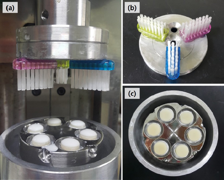
6개의 시편을 고정할 수 있도록 제작한 금형에 시편을 고정하고서 (Figure 1c), pin-on-disk 마모시험기 (Model KD-WT02, Kwangduck FA, Korea)를 이용하여 구강 환경에서 약 10년에 해당하는 100,000회 칫솔질(21)을 시행하였다 (Figure 1a). ISO 11609:2017 표준에 따라서 중경도 칫솔모를 갖는 칫솔 (Gum classic #311, Sunstar Americas Inc, USA) 3개를 원주상에 120도 간격으로 고정하여 사용하였고 (Figure 1b), 작용력은 2 N, 회전속도 100 rpm, 실온 약 27 ℃의 조건에서 칫솔질을 하였다(22). 칫솔질 과정에서 치약 25 g과 40 ml의 멸균증류수 (5 : 8 비율)를 혼합하여 사용하였고, 시편 위에 syringe로 치약 슬러리를 0.2 mL씩 주입하여 최소 두께 3 mm가 덮어지도록 하였다. 2분마다 혼합된 치약 슬러리를 0.2 mL씩 주입하였고, 칫솔은 5,000번의 칫솔질 횟수마다 교체하였다. 본 연구에 사용된 치약은 연마제 성분에 따라서 4종류로 분류하여 사용하였다 (Table 2).
3. 표면 형태 관찰
칫솔질 마모시험 후 시편을 초음파 세척기에서 10분간 멸균증류수로 세척한 후 상온에서 건조하여 사용하였다. 치약의 종류에 따른 시편의 표면 형태를 비교하기 위하여 광학현미경 (DM 2500, Leica Microsystems Co, Germany)과 주사전자현미경 (Scanning Electron Microscope; SEM, JSM-5900, Jeol Ltd, Japan)으로 관찰하였다.
4. 표면 거칠기
원자힘현미경 (Atomic force microscopy; AFM, Multimade-8, Bruker, USA)을 사용하여 칫솔질 마모시험 후 각 시편에서 3개의 다른 지점을 선택하여 표면 거칠기를 측정하였다. 표면 거칠기 3D 이미지와 평균 표면 거칠기 값 (Ra)은 fibers silicon nitride 팁과 접촉 모드를 사용하여 얻었고, 시편의 면적을 2 X 2 µm2로 하여 지점당 최소 2회 스캔하였다.
5. 통계 분석
측정된 표면 거칠기의 모든 값들은 유의수준 P=0.05에서 유의성을 검증하기 위해서 일원배치분산분석(One-way ANOVA test)을 시행하였고, 유의한 차이가 있는 변인에 대해서는 Tukey의 다중비교분석(Tukey’s multiple comparison test)으로 사후 검증을 하였다 (SPSS 22.0 software, SPSS Inc, USA). 유의성은 95% 신뢰 구간에서 5% 유의 수준으로 평가되었으며, P 값은 <0.05일 때 유의미한 것으로 간주되었다.
결과
Figure 2에서 Figure 4는 성분이 다른 치약들을 사용한 칫솔질 마모시험 후 미세 혼합형 및 나노필러 함유 복합레진의 표면을 보여주는 각 실험군들의 대표적인 광학 현미경과 SEM 이미지이다. Figure 2 (Z100 Restorative, Z1)와 Figure 3 (Filtek Z250, Z2)의 SEM 이미지들은 미세 혼합형 필러(microhybrid fillers)를 함유한 복합레진들의 표면 형태 변화를 관찰한 것으로 SEM 이미지 확인 결과, 대조군(Figure 2f, 3f)을 제외한 다른 실험군들의 표면에서는 상대적으로 큰 필러 입자들이 돌출되어 있고, 작은 분화구(crater) 형태의 결함들이 관찰되었다(Figure 2g-i, 3g-i). 제올라이트-M(Zeolite-M)을 함유한 메디안 치약으로 칫솔질 마모시험을 시행한 Z1, Z2 시편들에서는 레진기질의 선택적인 마모와 마이크로 필러들의 돌출 양상이 뚜렷하게 관찰되었다(Figure 2j, 3j). 광학 현미경으로 관찰한 Z1, Z2 시편들의 표면은 하이드록시아파타이트를 함유한 디오 치약, 탄산칼슘(CaCO3)을 함유한 페리오 치약 및 중탄산나트륨(sodium bicarbonate)을 함유한 Weleda 치약으로 마모시험을 시행한 후 스크래치 형태의 결함이 관찰되었고(Figure 2b-d, 3b-d), 제올라이트-M을 함유한 메디안 치약으로 마모시험을 시행한 Z1, Z2 시편들에서는 필러들의 돌출 양상이 관찰되었다(Figure 2e, 3e).
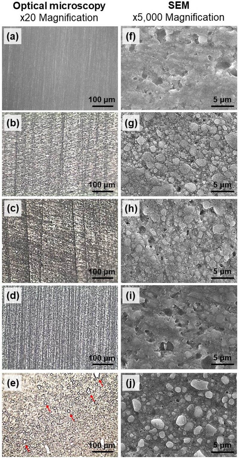
Optical microscopy (x20 magnification) and SEM images (x5,000 magnification) showing morphological changes on the surface of resin composite (Z100 Restorative, Z1) after toothbrushing with different types of toothpastes. The Z1N (a, f), the Z1D (b, g), the Z1P (c, h), the Z1W (d, i), and the Z1M groups (e, j). Red arrows indicate fillers.
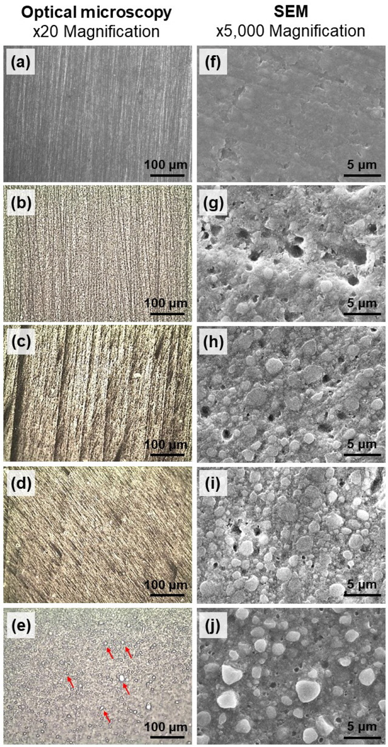
Optical microscopy (x20 magnification) and SEM images (x5,000 magnification) showing morphological changes on the surface of resin composite (Filtek Z250, Z2) after toothbrushing with different types of toothpastes. The Z2N (a, f), the Z2D (b, g), the Z2P (c, h), the Z2W (d, i), and the Z2M groups (e, j). Red arrows indicate fillers.
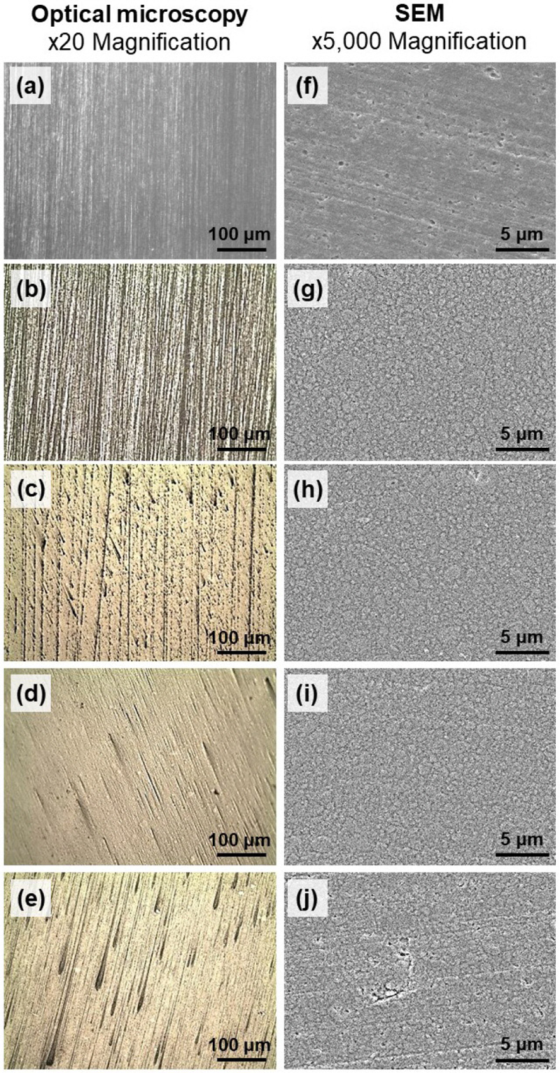
Optical microscopy (x20 magnification) and SEM images (x5,000 magnification) showing morphological changes on the surface of resin composite (Filtek Z350 XT, Z3) after toothbrushing with different types of toothpastes. The Z3N (a, f), the Z3D (b, g), the Z3P (c, h), the Z3W (d, i), and the Z3M groups (e, j).
Figure 4 (Filtek Z350 XT, Z3)의 SEM 이미지들은 나노필러(nano fillers)를 함유한 복합레진의 표면 형태 변화를 관찰한 것으로 대조군(Figure 4f)은 표면이 균일하고 변화가 거의 없어 매끄러워 보였다. 대조군을 제외한 다른 그룹들에서는 나노필러들의 돌출과 탈락 양상이 관찰되었으나 상대적으로 매끈한 표면을 보였고(Figure 4g-j), 메디안 치약으로 칫솔질 마모시험을 시행한 Z3 시편들에서는 불규칙적인 스크래치와 작은 결함들이 추가적으로 관찰되었다(Figure 4j). 광학 현미경으로 관찰한 Z3 시편들의 표면은 치약들로 마모시험을 시행한 후 불규칙한 스크래치 형태들이 관찰되었다(Figure 4b-e).
Figure 5에서 Figure 7은 성분이 다른 치약들을 사용한 칫솔질 마모시험 후 미세 혼합형 및 나노필러 함유 복합레진의 표면 거칠기 평균값(Ra, µm)과 AFM으로 스캔한 3D 이미지이다. 미세 혼합형 필러를 함유한 복합레진(Z100 Restorative, Z1)에서는 Z1M (0.208 ± 0.005) 그룹의 표면 거칠기 값이 유의하게(P<0.05) 가장 높았고(Figure 5a), 3D 이미지에서도 가장 큰 피크들(peaks)과 골들(troughs)을 가지고 있었다(Figure 5f). 대조군(Z1N, 0.038 ± 0.003)과 비교하여 다른 실험군들의 표면 거칠기 값은 유의하게 증가하였고(P<0.05), Z1P (0.095 ± 0.005)와 Z1W (0.107 ± 0.014) 그룹 사이에는 유의한 차이가 없었다(P>0.05).
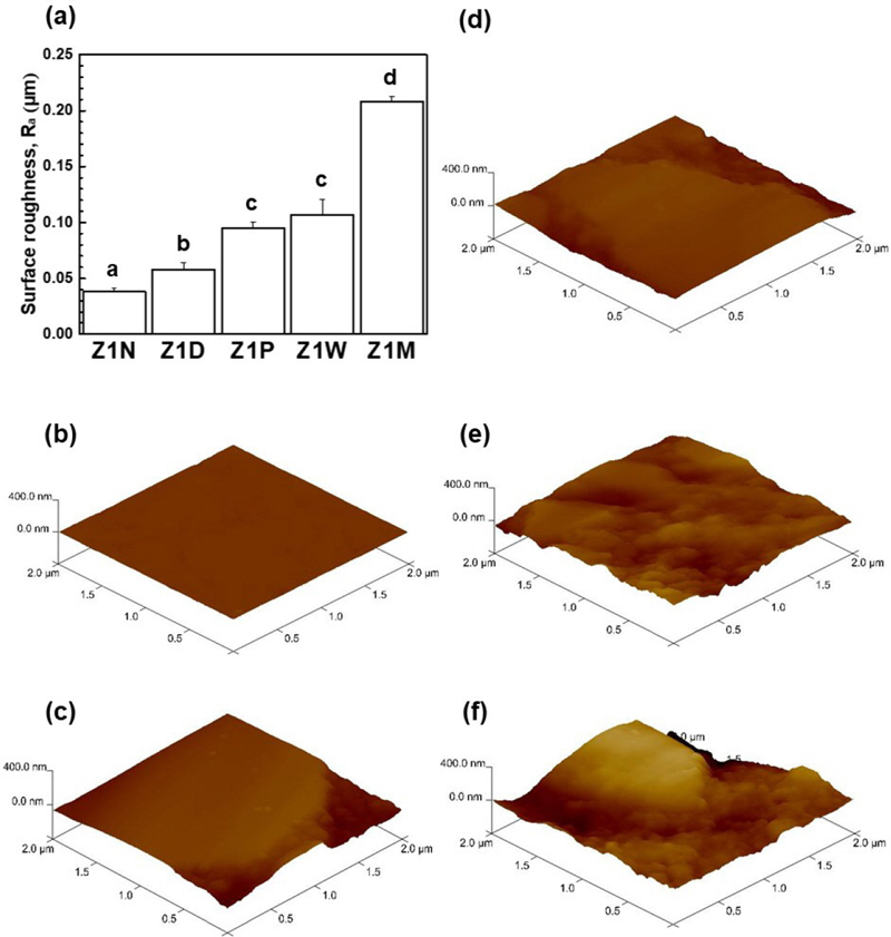
The resin composites' (Z100 Restorative, Z1) surface roughness values (Ra, μm) after toothbrushing with different types of toothpastes and 3D images scanned with AFM. (a) Means and standard deviations of surface roughness, (b) Z1N, (c) Z1D, (d) Z1P, (e) Z1W, and (f) Z1M groups.a-d Groups with different letters were significantly different (P<0.05).
미세 혼합형 필러를 함유한 복합레진(Filtek Z250, Z2)에서도 Z2M (0.266 ± 0.009) 그룹의 표면 거칠기 값이 유의하게(P<0.05) 가장 높았고(Figure 6a), 3D 이미지에서도 가장 큰 피크들과 골들을 가지고 있었다(Figure 6f). 대조군(Z2N, 0.038 ± 0.002)과 비교하여 다른 실험군들의 표면 거칠기 값은 유의하게 증가하였으나(P<0.05), Z2D (0.070 ± 0.005), Z2P (0.072 ± 0.005) 및 Z2W (0.078 ± 0.007) 그룹들 사이에서는 유의한 차이가 없었다(P>0.05).
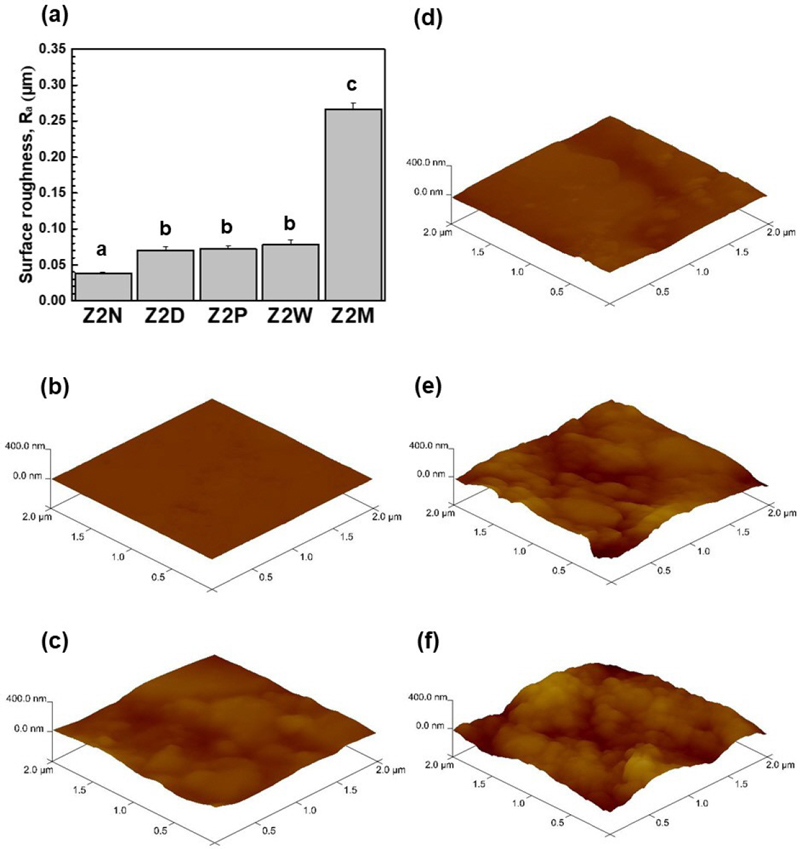
The resin composites' (Filtek Z250, Z2) surface roughness values (Ra, μm) after toothbrushing with different types of toothpastes and 3D images scanned with AFM. (a) Means and standard deviations of surface roughness, (b) Z2N, (c) Z2D, (d) Z2P, (e) Z2W, and (f) Z2M groups.a-c Groups with different letters were significantly different (P<0.05).
나노필러를 함유한 복합레진(Filtek Z350 XT, Z3)에서도 Z3M (0.090 ± 0.019) 그룹의 표면 거칠기 값이 유의하게(P<0.05) 가장 높았으나(Figure 7a), Z3W (0.089 ± 0.008) 그룹과는 유의한 차이를 보이지 않았다(P>0.05). 3D 이미지에서도 아주 낮은 피크들과 골들을 불규칙적으로 관찰할 수 있었다(Figure 7e, 7f). 대조군(Z3N, 0.046 ± 0.004), Z3D (0.057 ± 0.009), Z3P (0.072 ± 0.007) 그룹 순서대로 표면 거칠기 값이 유의하게 증가하였으나(P<0.05), 3D 이미지들에서는 거의 평평한 형태를 관찰하였다(Figure 7b-d).
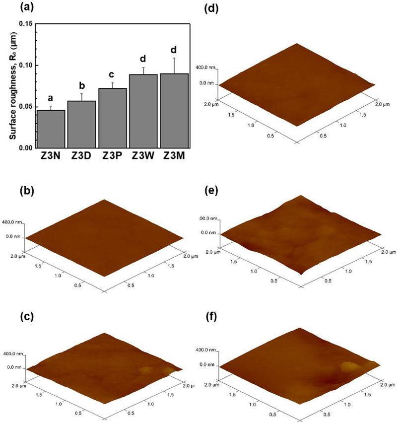
The resin composites' (Filtek Z350 XT, Z3) surface roughness values (Ra, μm) after toothbrushing with different types of toothpastes and 3D images scanned with AFM. (a) Means and standard deviations of surface roughness, (b) Z3N, (c) Z3D, (d) Z3P, (e) Z3W, and (f) Z3M groups.a-d Groups with different letters were significantly different (P<0.05).
고찰
임상적인 관점에서 복합레진의 표면 거칠기는 해당 재료의 내마모성, 착색, 치태 축적 및 이에 따른 치주 문제에 대한 민감성 측면으로 중요한 역할을 한다(23). 문헌들에 따르면 구강 조직 (치아, 치주조직)과 수복재료들을 청결하게 관리하기 위해서는 최소한의 마모도가 필요하다고 알려져 있다(24). 하지만, 치약 성분 중 연마제의 마모도 값이 매우 낮으면 수복재료의 표면에 착색과 치태 축적으로 구강 조직의 손상과 수복재료의 내구성에 문제가 생길 수 있다(23, 25).
구강 관리 및 위생을 위해서는 일반적으로 치약, 구강 세정액, 구강위생용품 (치실, 치간칫솔, 혀클리너 등)이 사용된다. 치약의 연마제 성분은 주로 실리카(silica), 수산화인회석(hydroxyapatite), 탄산칼슘(calcium carbonate), 알루미노규산나트륨(sodium aluminosilicate), 메타인산나트륨(sodium metaphosphate), 베이킹소다(중탄산나트륨, sodium Bicarbonate) 등의 수불용성(water-insoluble) 무기 분말로 구성되어 있다(26, 27). 여러 in vitro 연구에서는 치약에 포함된 연마제 성분이 복합레진의 색 안정성, 미세경도 및 표면 거칠기에 미치는 영향에 대해서 평가하였다(24, 28, 29). Roselino 등(24)에 따르면 미백치약의 연마제는 복합레진의 색상변화와 관련이 없고, 표면거칠기를 증가시킨다는 결론을 내렸다. Khamverdi 등(30)은 치약의 연마제 성분인 수화 실리카, 중탄산나트륨이 복합레진의 표면 미세경도를 감소시킨다고 하였고, 결과적으로 연마제 성분이 기질의 표면에 영향을 미친다고 하였다. 치약 연마제의 기능적인 중요성에도 불구하고, 치약에 포함되어 있는 다양한 연마제를 비교한 연구는 거의 없다(26).
따라서, 본 연구에서는 서로 다른 4종류의 연마제 성분을 포함한 치약을 사용하였고, 자세한 정보는 Table 2에 나열하였다. 또한, 칫솔질 마모시험을 시행하여 수복재료인 미세 혼합형 및 나노필러 복합레진의 표면 형태와 표면 거칠기를 관찰하여 내마모성을 평가하고자 하였다.
복합레진은 저작 중 연마성 음식물 슬러리의 이동이나 칫솔질과 같은 형태로 마모가 발생하여 필러 입자 사이의 레진기질 손실과 그에 따른 필러의 이탈 과정을 포함한다(31). 레진기질의 내마모성은 필러 입자의 함량 및 크기, 필러-레진기질의 결합정도, 수분 흡수로 인한 필러/레진기질의 균열과 관련 있는 것으로 보고되었다(32). 필러 입자의 함량 증가 및 크기의 감소, 필러-레진기질 간의 강한 결합력 등으로 복합레진의 내마모성이 향상된다(33). 저작이나 칫솔질 과정에서 복합레진의 레진기질은 필러 입자보다 먼저 제거되어 표면에 필러가 돌출되고, 상대적으로 큰 필러 입자를 함유한 혼합형, 미세 혼합형 복합레진은 나노필러 복합레진에 비해서 거칠고 불규칙한 표면을 갖게 된다(34). 이에 따른 문제점은 치태 축척으로 인한 이차우식증, 치은염증 유발과 수복재료의 착색 및 물리적 내구성의 저하 등이 있다(35).
지르코니아/실리카 함유 복합레진은 다른 성분(바륨, 유리, 석영 등)의 필러를 함유한 복합레진보다 우수한 내마모성을 나타냈다(36). 임상적 관점에서 볼 때, 더 많은 필러를 분해하는 유리 복합레진 대신 필러 입자가 레진기질에 확고하게 결합되는 지르코니아/실리카 함유 복합레진을 사용하는 것이 권장되며, 이는 내마모성에도 중요한 역할을 할 수 있다(33). 본 연구에서는 지르코니아/실리카 미세 혼합형 필러 복합레진과 나노필러 함유 복합레진 3종류를 사용하였다. 칫솔질 마모시험 후 미세 혼합형 복합레진들(Z100 Restorative와 Filtek Z250)의 표면 관찰 결과 상대적으로 큰 필러 입자들이 돌출되어 있고, 작은 분화구(crater) 형태의 결함들이 관찰되었다(Figure 2, 3). 제올라이트-M을 함유한 치약으로 마모시험을 시행한 그룹들의 표면 거칠기 값이 유의하게 가장 높았고(P<0.05)(Figure 5a, 6a), 레진기질의 선택적인 마모 및 마이크로 필러들의 돌출과 탈락 양상으로 가장 크고 깊은 결함들이 나타났다(Figure 2j, 3j). 제올라이트-M은 치석 형성에 관여하는 Ca2+와 같은 금속 양이온을 킬레이트화 제거하고, 외인성 착색을 방지하는 기능이 있다(37). 또한, 제올라이트-M은 수백 마이크로미터의 상대적으로 큰 입자를 가진 연마제로 높은 마모성과 관련이 있다고 하였고(38), 본 실험에서도 가장 높은 표면 거칠기 값과, 강렬한 표면 특징을 보이며 문헌과 일치하는 결과를 얻었다. 나노필러 함유 복합레진(Filtek Z350 XT)에서도 제올라이트-M으로 칫솔질 마모시험을 시행했을 때 표면 거칠기 값이 유의하게(P<0.05) 가장 높았으나, 치약 종류와 상관없이 표면 거칠기가 0.1 µm 미만으로 낮은 값들을 보였다(Figure 7a). 미세 혼합형 필러 복합레진들에 비해 표면이 균일하고 변화가 미미하여 매끄러워 보였다(Figure 7). Suzuki T 등(12)도 나노필러 소재 복합레진으로 칫솔질 마모시험을 시행하였고, 적당한 누적 마모 깊이와 균일하고 매끄러운 표면을 얻었다. 또한, SEM 결과에서 나노사이즈의 나노 클러스터들이 관찰되면서 마모 과정이 비교적 느리다고 발표하였다. Colak G 등(39)에 따르면, 치약으로 칫솔질 마모시험을 시행한 후 마이크로 하이브리드 복합레진의 표면 거칠기 값은 나노 복합레진의 값보다 높았고, 가장 낮은 표면 거칠기 값은 초나노 복합레진이라고 하였다. 문헌들에서는 칫솔질 후 필러 입자의 크기가 더 큰 복합레진에서 일반적으로 평균 표면 거칠기 값이 증가한다고 하였고(20, 40), 필러 크기가 클수록 필러가 더 많이 제거되어 수복물의 표면 거칠기가 증가된다고 하였다(41). 그러나, 필러의 형상, 필러 간의 거리, 필러 입자 간의 화학적 결합 및 중합 후의 전환 정도는 반드시 고려되어야 하는 요소이다(39).
학자들은 치약의 연마성과 수복재료의 표면 거칠기 사이에 물질 의존적 상호작용이 있다고 보고했다(42). 본 실험 결과 복합레진의 종류와 상관없이 제올라이트-M 함유 치약으로 칫솔질 마모시험을 한 후 표면 거칠기 값이 가장 높게 나타났으며, 하이드록시아파타이트 < 탄산칼슘 < 중탄산나트륨이 함유된 치약의 순서대로 표면 거칠기가 증가하였다(Figure 5-7). 4종의 치약들로 칫솔질 마모시험에 따른 표면 거칠기 값의 차이가 입증되었기 때문에 연마제 성분이 다른 치약들은 미세 혼합형 및 나노필러 복합레진의 표면 거칠기에 영향을 미치지 않으며, 치약과 복합레진의 필러 종류에 따른 표면 거칠기의 차이는 없을 것이라는 귀무가설은 기각되었다. 치약에 포함된 다양한 연마제 중에서 수산화인회석은 타액단백질과 결합하여 치태형성 억제, 치아 표면의 연마 효과 및 상아세관을 막아 지각과민증 완화에도 효과가 있다(43). 치아 마모의 억제를 위해서 비교적 연질인 탄산칼슘이 치약의 연마제로 사용되고 있으며(44), 탄산칼슘 입자는 레진기질이 빠르게 마모되는 것을 방지하고 필러가 기질에 더 오랫동안 유지되도록 한다(12). 중탄산나트륨은 연마율이 낮은 연마제 입자로 분류되고, 높은 용해도로 저마찰 특성을 가지고 있어 낮은 연마성을 보인다(26).
ISO 11609:2017 표준에 따라(22) 칫솔질 마모시험에 0.5-2.5 N의 하중을 적용해야하고, 본 연구에서는 2 N의 하중을 사용했다. 지르코니아/실리카 함유 복합레진(미세 혼합형 및 나노필러 복합레진)에 대한 칫솔과 치약의 영향을 조사한 결과 치약의 연마제 성분, 복합레진의 필러 입자의 크기가 복합레진의 표면 형태와 표면 거칠기에 영향을 주어 내마모성에 차이가 있음을 확인하였다. 칫솔질 마모시험 후 재료의 반경 약 0.8 µm의 표면 거칠기를 주의 깊게 해석해야 하며(12), 연구 결과 사용된 AFM의 팁 반경은 약 1~2 µm이므로 복합레진의 돌출된 필러 입자 사이의 좁은 공간을 측정하여 더욱 정확한 표면 거칠기 값을 표시할 수 있었다. 치약에 포함된 연마제의 종류에 상관없이 복합레진의 레진기질이 칫솔질의 결과로 마모되고 나머지 필러 입자가 거친 표면을 형성한다는 것을 보여주었다. 제올라이트-M 함유 치약으로 칫솔질한 경우 3종 복합레진의 표면 거칠기 값이 유의하게 증가하였다(P<0.05). 하지만, 나노사이즈의 필러가 함유된 복합레진 Filtek Z350 XT를 사용한 실험군들 간의 표면 거칠기 값의 차이는 크지 않았다. 따라서, 치약의 종류와 상관없이 표면 거칠기 값이 0.1 µm 미만이며, 표면 형태가 균일하고 매끄러운 복합레진 Filtek Z350 XT (나노필러 복합레진)가 상대적으로 더 높은 내마모성을 보였다.
현재 연구에는 몇 가지 한계점이 있다. 미생물군, 타액 순환, 온도 및 pH 변화와 같은 실제 구강 환경과 유사한 내마모성 시험이 필요할 것이며, 마모시험과 색상변화 및 경도 값 사이의 상관관계에 대한 추가 연구가 필요할 것이다. 또한, 생체 내 연구를 수행하기는 어렵지만 임상 실무 권고에 따른 추가 실험 연구와 다양한 분석 방법으로 복합레진 표면 변화의 메커니즘을 자세히 설명하는 연구가 필요하다.
결론
연구의 한계 내에서, 연마제 성분이 다른 4종의 치약들은 칫솔질 마모시험 후 복합레진에 다양한 표면 거칠기와 형태학적 변화를 보였다고 결론 내릴 수 있다. 제올라이트-M 함유 치약을 사용한 복합레진 그룹들에서 가장 높은 거칠기 값들이 관찰되었고(P<0.05), 하이드록시아파타이트 < 탄산칼슘 < 중탄산나트륨 함유 치약들의 순서대로 거칠기가 증가하였지만 큰 차이를 보이진 않았다. 칫솔질 마모시험 후 미세 혼합형 복합레진들(Z100 Restorative, Filtek Z250)의 표면 관찰 결과 상대적으로 큰 필러 입자들이 돌출되었고, 크고 깊은 결함들이 관찰되었다. 나노필러 복합레진(Filtek Z350 XT)은 치약의 종류와 상관없이 표면 거칠기 값이 0.1 µm 미만이었고, 표면 형태가 균일하고 매끄러웠다.
결과적으로, 미세 혼합형 복합레진에 비하여 나노필러 복합레진이 상대적으로 높은 내마모성을 보였으며, 칫솔질 내마모성의 특성은 임상에서 재료의 내구성을 나타낼 수 있다. 하지만, 칫솔모의 경도, 가해지는 압력, 칫솔질 기간 등은 복합레진의 내마모성에 더 중요한 역할을 할 수 있으므로 추가적인 연구가 필요할 것이다.
References
-
Tunc ES, Bayrak S, Guler AU, Tuloglu N. The effects of children's drinks on the color stability of various restorative materials. J Clin Pediatr Dent. 2009;34(2):147-50.
[https://doi.org/10.17796/jcpd.34.2.953q255621436788]

-
Nasim I, Neelakantan P, Sujeer R, Subbarao C. Color stability of microfilled, microhybrid and nanocomposite resins—an in vitro study. J Dent. 2010;38:137-42.
[https://doi.org/10.1016/j.jdent.2010.05.020]

-
Villalta P, Lu H, Okte Z, Garcia-Godoy F, Powers JM. Effects of staining and bleaching on color change of dental composite resins. J Prosthet Dent. 2006;95(2):137-42.
[https://doi.org/10.1016/j.prosdent.2005.11.019]

-
Schroeder T, da Silva PB, Basso GR, Franco MC, Maske TT, Cenci MS. Factors affecting the color stability and staining of esthetic restorations. Odontology. 2019;107:507-12.
[https://doi.org/10.1007/s10266-019-00421-x]

-
Szczesio-Wlodarczyk A, Sokolowski J, Kleczewska J, Bociong K. Ageing of dental composites based on methacrylate resins—a critical review of the causes and method of assessment. Polymers. 2020;12(4):882.
[https://doi.org/10.3390/polym12040882]

-
Dionysopoulos D, Gerasimidou O. Wear of contemporary dental composite resin restorations: a literature review. Restor Dent Endod. 2021;46(2).
[https://doi.org/10.5395/rde.2021.46.e18]

-
Azmy E, Al-Kholy MRZ, Fattouh M, Kenawi LMM, Helal MA. Impact of nanoparticles additions on the strength of dental composite resin. Int J Biomater. 2022;2022.
[https://doi.org/10.1155/2022/1165431]

-
Turssi C, Ferracane J, Vogel K. Filler features and their effects on wear and degree of conversion of particulate dental resin composites. Biomaterials. 2005;26(24):4932-7.
[https://doi.org/10.1016/j.biomaterials.2005.01.026]

-
Stawarczyk B, Özcan M, Trottmann A, Schmutz F, Roos M, Hämmerle C. Two-body wear rate of CAD/CAM resin blocks and their enamel antagonists. J Prosthet Dent. 2013;109(5):325-32.
[https://doi.org/10.1016/S0022-3913(13)60309-1]

-
Ayatollahi M, Yahya MY, Karimzadeh A, Nikkhooyifar M, Ayob A. Effects of temperature change and beverage on mechanical and tribological properties of dental restorative composites. Mater Sci Eng C Mater Biol Appl. 2015;54:69-75.
[https://doi.org/10.1016/j.msec.2015.05.004]

-
Nimri EM. Wear mechanisms and wear investigations of dental materials; a review of the literature. Arch Oral Den Res. 2015;2(3):1-7.
[https://doi.org/10.7243/2053-5775-3-4]

-
Suzuki T, Kyoizumi H, Finger WJ, Kanehira M, Endo T, Utterodt A, et al. Resistance of nanofill and nanohybrid resin composites to toothbrush abrasion with calcium carbonate slurry. Dent Mater J. 2009;28(6):708-16.
[https://doi.org/10.4012/dmj.28.708]

-
Kim SS, Cho JW, Lee CH. Gingivitis reducing effect of calcium glycerophosphate, cetylpyridinium chloride and dipotassium glycyrrhizate containing dentifrice. J Korean Soc Dent Hyg. 2017;17(6):983-92.
[https://doi.org/10.13065/jksdh.2017.17.06.983]

-
Kim YG, Park JH, Cho JW. Tooth whitening effects of manicure-type hydrogen peroxide tooth whitening gel. J Korean Acad Oral Health. 2019;43(4):191-5.
[https://doi.org/10.11149/jkaoh.2019.43.4.191]

- Turssi CP, Messias DC, Hara AT, Hughes N, Garcia-Godoy F. Brushing abrasion of dentin: Effect of diluents and dilution rate of toothpaste. Am J Dent. 2010;23(5):247.
-
Parry J, Harrington E, Rees GD, McNab R, Smith AJ. Control of brushing variables for the in vitro assessment of toothpaste abrasivity using a novel laboratory model. J Dent. 2008;36(2):117-24.
[https://doi.org/10.1016/j.jdent.2007.11.004]

-
Kumar S, Patnaik A, Bhat I. Factors influencing mechanical and wear performance of dental composite: a review. Materialwiss Werkstofftech. 2020;51(1):96-108.
[https://doi.org/10.1002/mawe.201900029]

-
Ruivo MA, Pacheco RR, Sebold M, Giannini M. Surface roughness and filler particles characterization of resin‐based composites. Microsc Res Tech. 2019;82(10):1756-67.
[https://doi.org/10.1002/jemt.23342]

- Cao L, Zhao X, Gong X, Zhao S. An in vitro investigation of wear resistance and hardness of composite resins. Int J Clin Exp Med. 2013;6(6):423.
-
Oliveira GUd, Mondelli RFL, Charantola Rodrigues M, Franco EB, Ishikiriama SK, Wang L. Impact of filler size and distribution on roughness and wear of composite resin after simulated toothbrushing. J Appl Oral Sci. 2012;20:510-6.
[https://doi.org/10.1590/S1678-77572012000500003]

-
Yuan JC-C, Barão VAR, Wee AG, Alfaro MF, Afshari FS, Sukotjo C. Effect of brushing and thermocycling on the shade and surface roughness of CAD-CAM ceramic restorations. J Prosthet Dent. 2018;119(6):1000-6.
[https://doi.org/10.1016/j.prosdent.2017.06.001]

-
O'Neill C, Kreplak L, Rueggeberg FA, Labrie D, Shimokawa CAK, Price RB. Effect of tooth brushing on gloss retention and surface roughness of five bulk‐fill resin composites. J Esthet Restor Dent. 2018;30(1):59-69.
[https://doi.org/10.1111/jerd.12350]

-
Dionysopoulos D, Papageorgiou S, Papadopoulos C, Davidopoulou S, Konstantinidis A, Tolidis K. Effect of whitening toothpastes with different active agents on the abrasive wear of dentin following tooth brushing simulation. J Func Biomater. 2023;14(5):268.
[https://doi.org/10.3390/jfb14050268]

-
Roselino LdMR, Rafaella TT, Cristian S, Ayodele AA, Carolina NFdA, Camila T, et al. Color stability and surface roughness of composite resins submitted to brushing with bleaching toothpastes: An in situ study. J Esthet Restor Dent. 2019;31(5):486-92.
[https://doi.org/10.1111/jerd.12495]

-
Roselino LdMR, Cruvinel DR, Chinelatti MA, Pires-de FdCP. Effect of brushing and accelerated ageing on color stability and surface roughness of composites. J Dent. 2013;41(5):54-61.
[https://doi.org/10.1016/j.jdent.2013.07.005]

-
Hara AT, Turssi CP. Baking soda as an abrasive in toothpastes: Mechanism of action and safety and effectiveness considerations. J Am Dent Assoc. 2017;148(11):S27-S33.
[https://doi.org/10.1016/j.adaj.2017.09.007]

-
Shuto T, Mine Y, Makihira S, Nikawa H, Wachi T, Kakimoto K. Alterations to titanium surface depending on the fluorides and abrasives in toothpaste. Materials. 2021;15(1):51.
[https://doi.org/10.3390/ma15010051]

-
Yilmaz MN, Gul P, Unal M, Turgut G. Effects of whitening toothpastes on the esthetic properties and surface roughness of a composite resin. J Oral Sci. 2021;63(4):320-5.
[https://doi.org/10.2334/josnusd.21-0044]

-
Kyoizumi H, Yamada J, Suzuki T, Kanehira M, Finger WJ, Sasaki K. Effects of toothbrush hardness on in vitro wear and roughness of composite resins. J Contemp Dent Pract. 2013;14(6):1137.
[https://doi.org/10.5005/jp-journals-10024-1464]

- Khamverdi Z, Kasraie S, Rezaei-Soufi L, Jebeli S. Comparison of the effects of two whitening toothpastes on microhardness of the enamel and a microhybride composite resin: an in vitro study. J Dent 2010;7(3):139.
-
Ishikiriama SK, Ordonez-Aguilera JF, Maenosono RM, Volú FLA, Mondelli RFL. Surface roughness and wear of resin cements after toothbrush abrasion. Braz Oral Res. 2014;29:1-5.
[https://doi.org/10.1590/1807-3107BOR-2015.vol29.0011]

-
Mandikos MN, McGivney GP, Davis E, Bush PJ, Carter JM. A comparison of the wear resistance and hardness of indirect composite resins. J Prosthet Dent. 2001;85(4):386-95.
[https://doi.org/10.1067/mpr.2001.114267]

- Say EC, Civelek A, Nobecourt A, Ersoy M, Guleryuz C. Wear and microhardness of different resin composite materials. Oper Dent. 2003;28(5):628-34.
-
Chen F, Sun L, Luo H, Yu P, Lin J. Influence of filler types on wear and surface hardness of composite resin restorations. J Appl Biomater Funct Mater. 2023;21:22808000231193524.
[https://doi.org/10.1177/22808000231193524]

-
Heintze S, Forjanic M, Ohmiti K, Rousson V. Surface deterioration of dental materials after simulated toothbrushing in relation to brushing time and load. Dent Mater. 2010;26(4):306-19.
[https://doi.org/10.1016/j.dental.2009.11.152]

- Gajapriya M, Somasundaram J, Geetha R. Fillers in composite resins-recent advances. Eur J Mol Clin Med. 2020;7(1):971-7.
- Ha WH, Ahn JH, You AR, Kim JH, Cho MJ, Shin SC. The interdental cleansing, stain removing and calculus deposit inhibition effect of toothpaste containing tetrasodium pyrophosphate and soft granule (Zeolite-M). Int J Clin Prev Dent. 2013;9(4):199-206.
-
Kim JH, Kim SY, TRUONG VM, Lee JW, Park YS. Is whitening toothpaste safe for dental health?: RDA-PE method. Dent Mater J. 2022;41(5):731-40.
[https://doi.org/10.4012/dmj.2021-303]

-
Colak G, Katirci G. In vitro evaluation of the effects of whitening toothpastes on the color and surface roughness of different composite resin materials. BMC Oral Health. 2023;23(1):580.
[https://doi.org/10.1186/s12903-023-03277-4]

-
Senawongse P, Pongprueksa P. Surface roughness of nanofill and nanohybrid resin composites after polishing and brushing. J Esthet Restor Dent. 2007;19(5):265-73.
[https://doi.org/10.1111/j.1708-8240.2007.00116.x]

- Ryba TM, Dunn WJ, Murchison DF. Surface roughness of various packable composites. Oper Dent. 2002;27(3):243-7.
-
Pires-de FdCP, Garcia LdFR, Roselino LdMR, Naves LZ. Color stability of silorane-based composites submitted to accelerated artificial ageing—an in situ study. J Dent. 2011;39:e18-e24.
[https://doi.org/10.1016/j.jdent.2011.03.003]

-
Kim SH, Park JB, Lee CW, Koo KT, Kim TI, Seol YJ, et al. The clinical effects of a hydroxyapatite containing toothpaste for dentine hypersensitivity. J Korean Acad Periodont. 2009;39(1):87-94.
[https://doi.org/10.5051/jkape.2009.39.1.87]

-
de Oliveira G, De Aveiro J, Pavone C, Marcantonio RAC. Influence of different toothpaste abrasives on the bristle end‐rounding quality of toothbrushes. Int J Dent Hyg. 2015;13(1):18-24.
[https://doi.org/10.1111/idh.12073]

