칼드웰룩 수술시 상악동구의 유지도구로서 풍선 도뇨관의 효용성
Abstract
The purpose of this study was to assess retrospectively the efficacy of balloon catheter inserted into maxillary sinus via natural ostium on Caldwell-Luc operation. From January 2008 to February 2013, the patients who were diagnosed as nonodontogenic maxillary sinusitis were included. Patency of ostium and thickness to sinus mucosa were examined in preoperative and postoperative computed tomography. Finally 8 patients were included and among them, catheters were inserted in 5 patients, and not in 3. Compared to preoperative state, ostial obstruction was resolved in 3 patients and degree of mucosal thickening was decreased in 4 patients in catheter insertion group. One patient showed increased mucosal thickening, and 2 patients did not showed any change. In the group without balloon catheter, ostial patency was observed in 2 patients preoperatively and 3 postoperatively. Degree of mucosal thickening was not changed in 2 patients and decreased in 1 patient. The patients underwent iliac bone graft to close the lateral window of sinus showed worse prognosis. Also, ostium maintained patency without balloon catheter in all patients with patent ostium preoperatively.
Keywords:
balloon catheter, Caldwell-Luc operation, maxillary sinusitis, natural ostium1. 서 론
만성 상악동염의 치료는 크게 세 가지의 목표를 가진다. 그것은 1) 상악동 점막을 건강하게 회복시키는 것; 2) 비강과 상악동을 개통시키는 것; 3) 2)의 방법으로 개통된 통로가 혈병의 섬유화로 인해 다시 폐쇄되지 않도록 유지하는 것이다(Korean Association of Oral and Maxillofacial Surgeons, 2013). 상악동 점막을 건강하게 회복시키기 위해 전통적으로 이용되던 방법은 칼드웰룩 수술 (Caldwell-Luc operation)로, 이것은 상악동의 측벽의 골절단을 통해 상악동내에 접근한 후 병적인 상악동 점막을 모두 제거하는 방법이다. 칼드웰룩 수술은 비강-상악동 개방술을 동반하게 되는데, 이 때 원래 비강과 상악동을 이어주는 상악동구 (natural ostium)보다 하방에 존재하는 하비갑개에 절개를 하는 하비도 골개창술 (inferior meatal antrostomy)을 사용해왔다 (Caldwell, 1893; Luc, 1897). 그리고 3)의 목적을 위해 개창부분을 바셀린 거즈를 이용하여 유지하는 방법이 사용되어 왔다. 하비도 골개창술은 중력에 의해 술 후 출혈이 하비도로 흘러리므로 비강을 통해 배출되기에 유리하며, 바셀린 거즈를 삽입하는 것은 상악동 내의 점막을 압박하여 술 후 지혈을 돕고 하비도 개창술 부위가 다시 폐쇄되는 것을 억제하리라는 판단에 근거한다.
그러나 최근 칼드웰룩 수술 후 장기간에 걸쳐 발생하는 술후 상악 낭종 (postoperative maxillary cyst)의 발생에 이러한 수술 방식이 영향을 준다고 보고되었다(Noyek, 1976). 하비도 개창술 부위는 수술 후 80%에서 다시 폐쇄되며(Korean Association of Oral and Maxillo- facial Surgeons, 2013), 이에 따라 상악동 내 잔존하던 호흡기 상피세포들이 비강과 개통되지 않는 골결손부를 형성하고, 결과적으로 낭종화된다는 것이다(Kubo, 1927, 1933). 이러한 합병증을 예방하기 위해 칼드웰룩 수술은 내시경을 이용한 기능적 내시경 상악동 수술로 대체되고 있다(Korean Association of Oral and Maxillofacial Surgeons, 2013). 그러나 상악동 내 감염원이 존재하거나 구강-상악동 누공이 존재하는 경우 내시경 상악동 수술의 효과에 한계가 있기 때문에(Kim & Duncavage, 2010), 여전히 칼드웰룩 수술은 널리 이용되고 있다.
또한 하비도 개창술은 자연적인 상악동구 (natural ostium)을 유지하거나, 상악동구가 폐쇄된 경우 구상돌기 절제술 (uncinectomy)을 이용하여 상악동구를 넓혀주는 중비도 개창술로 대체되었다(Arnes, Anke, & Mair, 1985; Davis, Templer, & LaMear, 1991). 바셀린거즈를 삽입하면 술 후 약 1주일 경 반드시 제거되어야 하는데, 이 때 거즈와 수술 부위가 유착되어 있을 수 있어 제거시 통증이 심하며, 재출혈의 우려가 있다(Kim & Duncavage, 2010; Kyriakos, 1977). 따라서 수술 후 상악동구를 유지하기 위해 collagen plug나 gelatin film 등의 여러 재료들의 사용이 제안되었다(Bartley & Morton, 1996; Catalano & Roffman, 2003; Sakamaki et al., 1998).
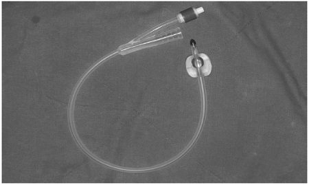
풍선 도뇨관 (balloon catheter, Fig. 1.)은 요도관을 통해 방광으로 삽입하는 용도로 고안된, 두 개의 분리된 채널로 이루어진 유연한 관이다. 원래는 스스로 소변을 보는 데 어려움이 있는 환자나 장시간의 수술을 하는 환자, 소변량을 감시해야 하는 중환자에서 요도에 삽입하는 용도로 사용되어 왔다. 두 개의 채널 중 하나의 내강은 양쪽 끝에서 열려있으며, 다른 내강은 안쪽 끝은 풍선처럼 부풀어오를 수 있고, 바깥쪽 끝은 공기나 물을 주입할 수 있는 밸브로 이루어져 있다. 풍선처럼 부풀어 오를 수 있는 안쪽 끝은 방광 내에 위치하게 되어 관이 바깥으로 미끄러져 나오지 않게 한다. 이것은 일반적으로 실리콘 러버나 러버로 만들어진다(Park et al., 2011). 이러한 도뇨관의 형태적 특성에 주목하여, 바셀린 거즈를 대체하는 수단으로써 상악동수술 후 비강을 통해 도뇨관의 balloon을 상악동 내에 삽입하면, 술 후 감염 방지 및 지혈 효과를 얻을 수 있으며, 제거시 통증과 재출혈의 위험이 감소한다는 보고들이 있었다(Lindorf, 1984; Sakamaki et al., 1998; Y et al., 2002). 그러나 이러한 방법들은 내시경을 이용한 기능적 상악동수술에 응용되고 있으며 아직 칼드웰룩 수술에서 술 후 상악동구의 개통을 유지하기 위해 사용된 결과는 보고된 바 없다. 저자들은 칼드웰룩 수술 후 상악동구를 통해 상악동 내에 삽입된 도뇨관이 상악동염의 치료에 대해 미치는 영향에 대해 확인하고자 하였다.
2. 재료 및 방법
이 후향적 연구는 원광대학교 치과병원 임상시험심사위원회의 승인 하에 진행되었다(WKDIRB201502-01). 2008년 1월부터 2013년 2월까지 원광대학교 치과대학병원 구강악안면외과에 내원하여 상악동염으로 진단된 환자들 중, 수술 전에 컴퓨터 단층촬영 영상과 파노라마 방사선사진이 촬영되었고, 술 후 1개월 이상 경과 관찰시에 컴퓨터 단층촬영 영상이 가능했던 환자들을 대상으로 하였다. 내원 이전에 상악동염으로 칼드웰룩 수술이나 내시경 상악동 수술을 받은 병력이 있는 환자들과, 내원 당시 치아의 감염과 연관된 상악동염으로 치아 치료를 선행해야 했던 환자들은 연구대상에서 제외하였다.
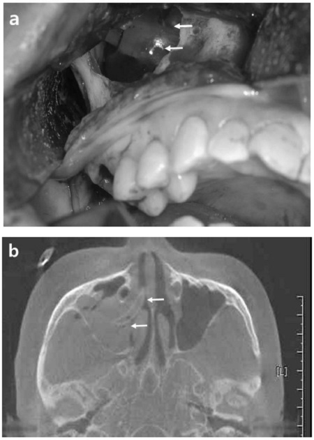
칼드웰룩 수술을 위해, 상악동의 측벽골을 절단하여 상악동에 접근하여 내부의 병적인 점막을 제거하였다. 비강과 상악동구를 통해 풍선 도뇨관을 상악동 내에 삽입한 후, 풍선에 식염수를 주입하여 상악동 부피에 맞게 확장시켰다(Fig. 2.). 골절단되었던 상악동 측벽은 골결손의 회복 없이 구강점막으로 덮거나, 장골이식편을 금속판과 나사못으로 고정하여 회복하여 주었다. 항생제는 술 전 상악동염의 증상에 따라 투약을 시작하여 수술 후 약 2주일까지 유지하였다. 수술 전 풍선 도뇨관은 수술 1주 후에 제거하였다.

Balloon catheter (white arrows) inserted into maxillary sinus. (a) Intraoperative photography shows balloon catheter inflated in maxillary sinus via removed lateral window. (b) Immediate postoperative computed tomography shows radiopaque indentation of balloon catheter inserted via sinus ostium.
환자들의 의무기록을 검토하여 환자의 연령, 성별, 상악동염 발생부위, 수술 전 후의 임상 증상, 수술 방법, 그리고 수술 중 풍선 도뇨관을 원래 상악동구로 삽입하였는지를 확인하였다. 도뇨관의 사용 여부에 따라 환자를 두 집단으로 분류하였으며, 컴퓨터 단층촬영 영상을 이용해 수술 전과 1개월 이상 경과후의 상악동구의 개방여부와 상악동 점막의 비후된 두께를 측정하였다. 상악동 점막의 비후 정도는, 상악동구가 관찰되는 컴퓨터 단층촬영영상의 관상단면상에서 상악동의 전체 높이에 대해 점막이 채운 높이를 각각 25%, 50%, 75%, 그리고 100%로 계산하였다.
3. 결 과
상악동염으로 인해 칼드웰룩 수술을 받은 환자들 중 이전 상악동염 수술 병력이 없고, 발치 등의 치아 치료가 필요 없었던 16명의 환자들 중, 수술 후 1개월 이상 경과관찰 시에 컴퓨터 단층 영상을 촬영한 환자는 8명이었다(표 1). 이 환자들의 평균 연령은 47.9세였고, 이 중 여성이 3명, 남성이 5명이었다. 상악동염이 좌측 상악동에 발생한 경우가 3증례, 우측에 발생한 경우가 5증례였다. 모든 환자에서 칼드웰룩 수술이 시행되었으며, 5명의 환자에서는 장골이식을 이용한 상악골 측벽의 즉시 회복이 동시에 이루어졌다.
전체 8명의 환자들 중 상악동구의 폐쇄가 술 전 5명에서 관찰되었고, 술 후에는 1명에서 관찰되었다. 점막비후의 정도는 술 전 3명에서 50%, 1명에서 75%, 4명에서 100%였으며, 술 후에는 4명에서 50%, 2명에서 75%, 2명에서 100%였다(표 1). 5증례에서 풍선 도뇨관을 삽입하였고(1군), 3증례에서는 삽입하지 않았다(2군).
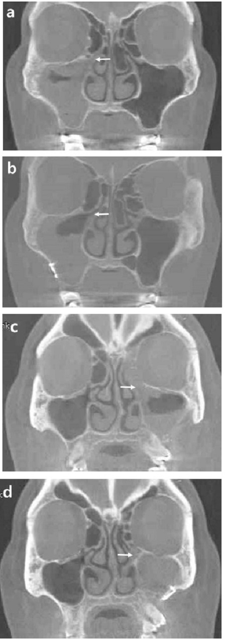
풍선 도뇨관을 삽입한 집단 중 상악동구가 폐쇄가 술 전 4명에서 관찰되었으나 수술 후에는 1명에서 관찰되어, 술 후 상악동구가 개방되는 경향을 보였다. 이 집단에서 상악동 점막의 비후 정도는 2증례에서는 수술 전(100%)보다 수술 후(75%)에 감소했다. 2증례에서는 점막비후의 변화가 없었고(100%; 50%), 한 명에서는 점막비후가 술 전(75%)보다 술 후(100%)에 증가했다(Fig. 3.). 풍선 도뇨관을 삽입한 환자들은 수술 후 풍선도뇨관 삽입에 대해 특기할만한 불편감을 호소하지 않았으며, 풍선 도뇨관의 삽입 여부에 관계없이 약간의 비충혈과 비출혈을 종종 호소하였다.

The patients with balloon catheter insertion on Caldwell-Luc operation. (a) preoperative and (b) postoperative computed tomography shows ostium obstruction (white arrows) was relieved after operation and mucosal thickening was decreased on right maxillary sinus. (c) preoperative and (d) posteoperative imaging shows ostium was still obstructed (white arrows) and mucosal thickening increased on left maxillary sinus.
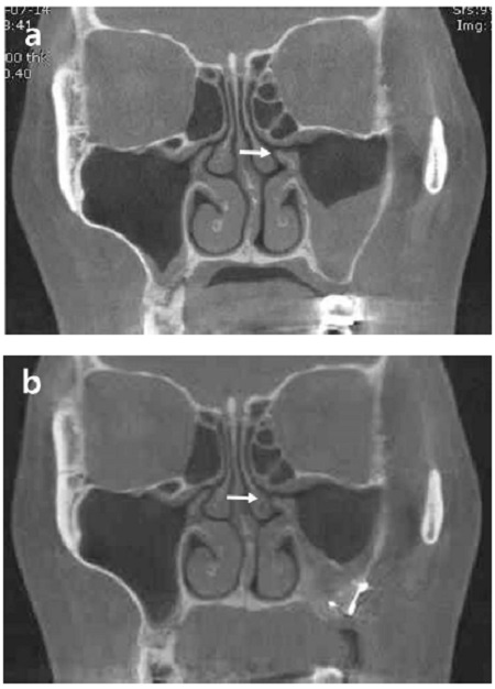
도뇨관을 삽입하지 않은 집단에서 상악동구의 폐쇄는 술 전 1증례에서 보였으며, 수술 후에는 모든 증례에서 상악동구 개방이 관찰되었다. 상악동 점막 비후 정도는 1명에서 술 후 감소(100%→50%)한 것이 관찰되었고, 2명의 환자에서는 변화가 없었다(50%, Fig. 4.).
4. 고 찰
풍선 도뇨관을 삽입한 후 1개월 이상 경과관찰 하면서 촬영한 컴퓨터 단층촬영 영상에서 상악동구는 거의 모든 환자에서 개방된 상태로 유지되었다. 수술 후 상악동구의 개방성과 점막 비후의 측면에서 모두 개선이 없거나 악화된 경우는 모두 4증례였는데, 이 네 증례는 모두 골이식을 동반한 증례였다. 칼드웰룩 수술 시에 절제되었던 상악동 측벽골을 골로 회복하지 않는 경우, 협측의 연조직이 상악동내로 함입되어 상악동의 부피를 감소시키고 상악동 내 점액의 배액과 환기를 방해한다고 알려져 있다(Korean Association of Oral and Maxillofacial Surgeons, 2013). 따라서 측벽골을 회복하기 위한 골이식술이 병행되어 왔다. 이 연구 결과, 골이식을 한 증례는 모두 6증례로, 이 중 2증례에서만 술 후 상악동구의 개방과 점막비후의 개선이 관찰되었다. 골이식을 한 경우에는 이식편의 생착 과정에서 염증반응이 호발할 수 있다고 알려져 있으며(Baek et al., 2010), 상악동 측벽의 골절단 및 수술 후에 절단된 골을 재식하여 고정한 경우에도 술 후 염증, 이식골의 이물반응, 혹은 상악동염이 재발될 수 있다는 보고가 있다(Kurokawa, Takeda, Yamashita, Nakamura, & Takahashi, 2002). 이러한 연구 결과들을 고려했을 때, 상악동염의 수술 시에 동시 골이식을 고려한다면 골이식 실패에 따른 감염 가능성을 고려한 접근이 필요하다고 생각된다.
또한 상악동구가 술 전에 개방되어 있던 환자에서는 칼드웰룩 수술시에 풍선 도뇨관을 상악동내 삽입하지 않아도 술 후 상악동구가 개방된 상태를 유지할 수 있었다. 많은 저자들이 하비도 개창술보다 원래의 상악동구를 확장하는 중비도 개창술 후 절개부위가 개통상태를 유지할 가능성이 높다고 주장하고 있다(Arnes et al., 1985; Davis et al., 1991; Kenned et al., 1987). 그러므로 원래 상악동구가 개통되어 있던 환자에서는 칼드웰룩 수술 후 혈병과 섬유화, 그리고 흉터에 의해 일시적으로 상악동구가 폐쇄되더라도 풍선 도뇨관의 삽입 없이 다시 개통될 수 있다고 가정할 수 있고, 본 연구에서도 술 전 상악동구가 개통되어 있던 두 명의 환자는 풍선 도뇨관 삽입 없이도 술 후 다시 상악동구가 개통상태로 회복되었음을 확인하였다.
이 연구에 포함된 3명의 환자는 내원 당시 상악 구치부 치아들의 치통을 호소했다. 상악동과 연관된 치아의 치주염이나 치근단염과 같은 치성 원인이 발견되어 치아 치료를 받아야 했던 환자들을 연구에서 제외했으나, 이 환자들에서는 파노라마 방사선 사진과 탐침, 생활력 검사를 통해 치성 원인을 배제하였기 때문에 이 연구에 포함하였다. 상악동염은 상악 구치부 치아의 동통으로 나타날 수 있으며(Korean Association of Oral and Maxillofacial Surgeons, 2013), 따라서 상악동과 인접한 상악 구치부 치아의 통증을 주소로 내원한 환자의 경우 상악동염을 의심할 수 있다.
그러나 본 연구는 치과 임상과의 특성상, 많은 상악동염 환자가 있음에도 불구하고 대다수가 치성 원인이 존재하기 때문에, 치성 원인을 배제한 후 연구에 포함된 환자가 8명에 불과했다는 한계가 있다. 향후 풍선 도뇨관을 이용한 상악동구 개통술의 효용성을 분석하기 위해서는 더 많은 환자를 확보하고, 더 장기간의 경과 관찰 결과를 이용한 연구가 필요할 것으로 생각된다. 또한 이번 연구에서 술 후 상악동염의 조절을 어렵게 할 가능성으로 제시된 골이식이 상악동염의 수술적 치료 후 경과에 어떤 영향을 미치는 지에 대해서도 통계적 분석을 통한 평가가 필요하리라 사료된다.
5. 결 론
본 연구에서는 칼드웰 수술 후 상악동구를 통해 상악동 내에 풍선 도뇨관의 삽입 여부에 따라 상악동염의 치료 결과의 차이를 비교 분석하였다. 본 연구에서는 제한된 환자 수로 다음과 같은 결론을 얻을 수 있었다.
- 1) 상악동염의 수술 시에, 풍선 도뇨관의 삽입여부에 관계없이 동시 골이식을 고려한다면 골이식 실패에 따른 감염 가능성을 고려해야 한다.
- 2) 제한된 환자 수로 인해 풍선 도뇨관의 삽입이 상악동염의 개선시킨다는 명확한 증거는 찾을 수 없었다.
- 3) 향후 풍선 도뇨관을 이용한 상악동구 개통술의 효용성을 분석하기 위해 더 많은 환자에서 장기간의 경과 관찰 연구가 필요하다.
Acknowledgments
* 이 연구는 2013년 원광대학교 교내연구비 지원에 의해 수행되었음.
참 고 문 헌
- Arnes, E., Anke, IM., & Mair, IW., (1985), A comparison between middle and inferior meatal antrostomy in the treatment of chronic maxillary sinus infection, Rhinology, 23(1), p65-69.
-
Baek, CH., Park, JH., Kim, GJ., Hong, JR., Kim, CS., & Paeng, JY., (2010), Comparison of healing pattern with or without bone graft after odontogenic cyst enucleation, J Korean Assoc Oral Maxillofac Surg, 36, p515-519.
[https://doi.org/10.5125/jkaoms.2010.36.6.515]

-
Bartley, J., & Morton, R., (1996), Complications of the Caldwell-Luc operation and how to avoid them, Aust N Z J Surg, 66(2), p119.
[https://doi.org/10.1111/j.1445-2197.1996.tb01128.x]

- Caldwell, GW., (1893), Disease of the accesory sinuses of the nose and an improved metho of treatment for suppuration of the maxillary antrum, NY Med J, 58, p526-531.
-
Catalano, PJ., & Roffman, EJ., (2003), Evaluation of middle meatal stenting after minimally invasive sinus techniques (MIST), Otolaryngology--Head and Neck Surgery, 128(6), p875-881.
[https://doi.org/10.1016/S0194-5998(03)00469-8]

- Davis, WE., Templer, JW., & LaMear, WR., (1991), Patency rate of endoscopic middle meatus antrostomy, The Laryngoscope, 101, p416-420.
-
Kenned, DW., Zinreich, SJ., Kuhn, F., Shaalan, H., Naclerio, R., & Loch, E., (1987), Endoscopic middle meatal antrostomy: theory, technique, and patency, The Laryngoscope, 97(S43), p1-9.
[https://doi.org/10.1288/00005537-198708002-00001]

-
Kim, Esther, & Duncavage, James, A., (2010), Prevention and management of complications in maxillary sinus surgery, Otolaryngologic Clinics of North America, 43(4), p865-873.
[https://doi.org/10.1016/j.otc.2010.04.011]

- Korean Association of Oral and Maxillofacial Surgeons (Ed.), (2013), Textbook of Oral and Maxillofacial Surgery, (3rd Edition ed.), Seoul, Dental and Medication Publishing Co.
- Kubo, I., (1927), Cheek cyst appeared after radical operation of maxillary sinusitis, Japanese Journal of Otolaryngology, 33, p896.
- Kubo, I., (1933), Postoperative wangenzyste, Z Otol Tokyo, 39, p1831-1845.
-
Kurokawa, H., Takeda, S., Yamashita, Y., Nakamura, T., & Takahashi, T., (2002), Evaluation of a modified method for maxillary sinus surgeryreimplantation of the anterior bony wall of the maxillary sinus, Asian Journal of Oral and Maxillofacial Surgery, 14(3), p144-147.
[https://doi.org/10.1016/S0915-6992(02)80035-8]

- Kyriakos, M., (1977), Myospherulosis of the paranasal sinuses, nose and middle ear. A possible iatrogenic disease, American journal of clinical pathology, 67(2), p118-130.
-
Lindorf, HH., (1984), Osteoplastic surgery of the sinus maxillaris-the “Bone lid”-method, Journal of Maxillofacial Surgery, 12, p271-276.
[https://doi.org/10.1016/S0301-0503(84)80258-1]

- Luc, H., (1897), Une nouvelle méthode opératoire pour la cure radicale et rapide de l’empyème chronique du sinus maxillaire, Arch Int Laryngol Otol Rhinol, 10, p273-285.
- Noyek, AM., (1976), Radiology of the maxillary sinus after Caldwell-Luc surgery, Otolaryngol Clin North Am, 9(1), p135-151.
-
Park, C. H., Kim, H. S., Lee, J. H., Hong, S. M., Kim, S. K., Ko, Y. G., Kang, T. H., (2011), A balloon dilatation technique for the treatment of intramaxillary lesions using a Foley catheter in chronic maxillary sinusitis, Am J Otolaryngol, 32(4), p304-307.
[https://doi.org/10.1016/j.amjoto.2010.07.001]

-
Sakamaki, H., Kujiraoka, H., Seki, M., Nema, T., Tsurumi, T., & Akiba, M., (1998), A comparative study of the usefulness of maxillary sinus tampons, Japanese Journal oF Oral and Maxillofacial Surgery, 44, p354-356.
[https://doi.org/10.5794/jjoms.44.354]

-
Y. Hamada, T. Kondoh, E. Imamura, H. Kanemura, H. Sekiya, K. Nakaoka, K. Seto, (2002), A unique balloon catheter for maxillary sinus surgery, Asian Journal of Oral and Maxillofacial Surgery, 14(4), p215-218.
[https://doi.org/10.1016/S0915-6992(02)80006-1]


