Bone formation of the porous layer formed of Ti-Ag mesh for GBR membrane applications

초록
본 연구의 목적은 마이크로 표면 위에 나노 단위의 다공성 구조를 갖는 티타늄-은 메쉬를 제작하고 메쉬의 골 형성능을 평가하는데 있다. 우선 시편 제작을 위해 cp-Ti에 비해 부식저항성이 뛰어난 티타늄-은 합금을 은 2.0 at%을 첨가시켜 제작하였다. 그리고 표면에 다공성 구조를 형성하기 위해 알루미나 입자를 사용하여 시편을 블라스팅 처리한 뒤 0.5% 불산 전해액에서 60분 동안 20 V 전압을 가하여 양극산화 처리하였다. 시편의 표면 형태는 주사전자현미경을 통해 관찰하였다. 골 형성능을 평가하기 위해 토끼의 두개골을 천공한 뒤 파절편을 제거하고 그 위에 시편을 식립하였다. 각각 2주와 4주 뒤 메쉬 주위의 골 조직 형태를 3D 스캔 이미지로 관찰하였고 생성된 골의 부피를 측정하였다. 시편 표면 형상 관찰 결과 마이크로 단위의 반구 형태 구조 위에 나노 단위의 다공성 구조를 관찰할 수 있었고, 골 형성능 결과에서는 대조군으로 메쉬를 식립하지 않은 경우에서 보다 티타늄-은 합금 메쉬를 식립한 경우에서 2주와 4주 모두 유의하게 더 많은 골 형성량을 보였다(p<0.05). 따라서 이 연구 결과로 미루어 보아 티타늄-은 합금에 마이크로 표면 위에 나노 다공성 구조를 형성할 수 있었으며 골 형성능 역시 우수하여 골유도 재생술을 위한 메쉬로서 유용하게 사용할 수 있을 것이라고 사료된다.
Keywords:
GBR (Guided Bone Regeneration) membrane, Micro surface-nano porous layer, Osseointegration, Ti–Ag alloyⅠ. INTRODUCTION
Restoration of bone is often required after dental and oral surgical procedures. Treatment of cysts, tumors, and fractures of the jaw can result in bone defects. Such defects must be repaired with bone grafts or bone substitutes to ensure good structural and functional outcomes (Nagao et al., 2002).
An ideal biomaterial should induce rapid, predictable and controlled healing of the host tissues (Brunski et al, 2000). The surface of an implant plays an important role in determining the biocompatibility and ultimately integration because it is in direct contact with the tissues (Nanci et al., 1998: Puleo and Nanci, 1999). It should be important for application of guided bone regeneration (GBR) membrane.
Various surface textures have been created and used successfully to influence the cell and tissue responses (Boyan et al., 1998). Among these, microtextured surfaces have been proposed as an important tool for establishing an advantageous, three-dimensional environment for osteogenic cells (Matsuzaka et al., 2003). Experimental data and the preliminary results from clinical trials revealed an effect on bone formation, at least during the initial stages of osseointegration (Park et al., 2001: Cochran et al., 2002).
It was demonstrated that different cell types respond to nanotopography (Curtis and Wilkinson, 1999). Cell/matrix/ substrate interactions associated with cell signaling can occur on the nanoscale. Such signaling regulates cell attachment, spreading, migration, differentiation, and gene expression (Schneider et al., 2001).
Ti-Ag alloys are considered to have enhanced corrosion resistance due to the formation of a stable passive oxide film. The addition of silver to titanium is believed to improve the corrosion resistance because of the easy formation of titanium oxide. In other words, silver, which has a much higher electromotive force than titanium, first facilitates the dissolution of titanium in artificial saliva, which then forms a stable titanium oxide film over the Ti-Ag alloy surface (Oh et al, 2005; Shim et al., 2005; Shim et al., 2006).
The aim of this study was to evaluate biocompatibility and osseointegration of the porous layer formed with both micro structure and nano structure on Ti-Ag mesh for GBR membrane applications.
Ⅱ. MATERIALS AND METHODS
1. Specimen preparation
Ti was used as commercially pure titanium (Hyundai, Grade 2). Sponge titanium (purity 99.99%) and granular silver (Yesbio, purity 99.99%) were used as the raw materials for alloy production. Titanium-silver alloys containing silver (2.0 at%) were designed. After weighing the designed composition, a 30 g quantity was melted four times in an arc melter to promote chemical homogeneity. The chamber was evacuated to 0.66 Pa and high-purity argon gas was introduced until the pressure reached 26 kPa before melting. After melting, the alloys were arc melted, homogenized at 950℃ for 72 h, and hot-rolled to 1 mm thickness. The alloys were then solution annealed at 950℃ for 1 h and quenched in water.
cp-Ti and Ti-Ag alloy were sectioned using a cutting machine (10×10×0.15 mm) and made holes by using an engine lathe. The hole diameter was 0.7 mm.
2. Surface Treatment
In order to obtain a micro-metric rough surface, the specimens were grit-blasted for 20 seconds using a 110 ㎛ aluminum oxide grit (Renfert, Germany) at a pressure of 4 bar and a distance of 10 mm from the sandblaster nozzle tip (2 mm in diameter). After grit-blasting, the specimens were washed for 10 minutes in distilled water using an ultrasonic cleaner. A nano-metric rough surface was obtained by anodizing the grit blasted specimens at 20 V for 60 min, in a 0.5% HF aqueous solution at room temperature using a DC power supply (Genesys 600-2.6, Densi-Lambda, japan). After anodic oxidation, the specimens were rinsed in distilled water. The specimens were annealed at 350 ˚C for 3 hours.
3. Surface Characteristics
The simple machined and porous surfaces were coated uniformly with gold for the electric conductivity, and their surface morphology was observed using scanning electron microscopy (SEM, Model S-800, Hitachi, Japan).
4. Histomorphometrical evaluation (Bone-metal contact)
The in vivo study was performed under the guidelines of the Animal Care and Use Committee, Yonsei Medical Center, Seoul, Korea. Twelve adult male rabbits weighting 3.20 ± 0.25 ㎏ were used (Samtako, Seoul, Korea). A midline incision was performed down to the bone surface of the calvarial bone, and a skin periosteum flap was raised to expose the calvarial bone on both sides of the midline. The outer cortical layer of calvarium bone was removed by trephine burr, and a 8 mm diameter cortical bone defect was made in each side of the calvarial bone; thus, a total of two bone defects were made in each animal (Figure 1). The meshes or without mesh on hole were covered for healing. Six of the twelve animals were euthanized after a healing period of 2 weeks and the other six animals after 4 weeks for histomorphometrical evaluation. Computed tomography (CT) images of the calvarial bone were taken for each rabbit (Model skyscan 1076, Skyscan, Belgium). CT analysis was used to analyze differences in the percentage areas of newly mineralized bone after 2 weeks and 4 weeks of healing.

(a) A cutaneous flap was demarcated, and then the periosteum was removed and the calvarial bone exposed on both sides of the midline. On each side, a circle 8 mm in diameter was made with a trephine bur. (b) Titanium meshes either with or without surface treatment of between cp-Ti and Ti-Ag mesh placed in the bone and the mesh fixed by sewing.
Ⅲ. RESULTS AND DISCUSSION
1. Surface Characteristics
Figure 2 shows a photomicrograph of porous layer mesh. There is made by grit-blasting and anodic oxidation. The surface consisted of nano-metric pores on the micro-metric surface with a hemisphere shape. The diameter of the micro-metric rough surface ranged from 1 to 10 ㎛, and micro-metric pores were separated irrelgularly on the mesh. The diameter of the nano-metric pores ranged from 40 to 100 nm, and nano-metric pores were well separated uniformly over the micro-metric pores. The initial attachment of cells is important for implantation of dental implant. The implant surface with micrometer-ordered pore structure increases the surface area and consequently increases the osseointegration efficiency(Abron et al., 2001). The adhesion of cells such as osteoblasts is a prerequisite to subsequent cell functions such as the synthesis of extracellular matrix proteins and the formation of mineral deposits. The introduction of nanostructured ceramics significantly improves osteoblast adhesion (Webster et al., 2000; Webster et al., 2001b)
2. Histomorphometrical evaluation (Bone-metal contact
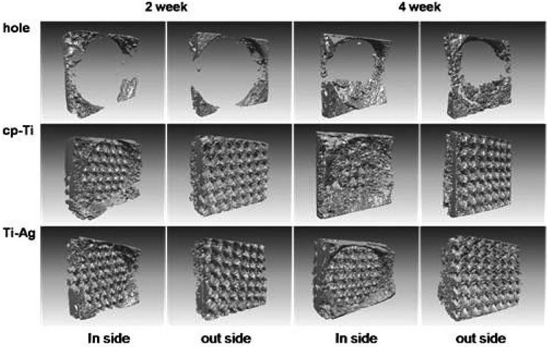
Figure 3 shows newly regenerated bone formed after a mesh implanted to calvaria bone. In the hole without mesh, regenerated bone didn’t entirely fill for 4 weeks. But, the newly formed bone with mesh was more formed than without mesh.

Computed tomography (CT) images for histomorphometrical evaluation of implanted meshes (10×10×0.15 mm). Newly generated bone tissue formed on the mesh was observed by 3-D scanning image. (Gray: newly generated bone, blue: mesh, in side: inner of the mesh, outside: outer of the mesh) the images were observed about implantation periods between inside and outside each of 2 weeks and 4 weeks.
Figure 4 shows the volume of mineralized bone in newly generated tissue on the mesh. Ti-Ag mesh produced more bone than the hole case without mesh in rabbit calvaria experiment (P<0.05). And in containing mesh on hole, we observed that results of 4 weeks were occurred newly bone better than 2 weeks. We thought a mesh might affect to formation of regenerated bone. We observed newly formed bone at the outer mesh. In this case, growth factor might transfer from hole of mesh.

Volume of mineralized bone in newly generated tissue on the mesh (White graphs are 2 weeks result and grey graphs are 4 weeks result).
The excellent biocompatibility of titanium and the easy handling of the titanium micro-mesh systems allowed their application for three-dimensional reconstruction of large bony defects. The most likely hypothesis that can explain the apparent benefits of the mesh lies in its probable protective effect during the healing time following bone grafting, as already found with non-resorbable membranes (Antoun et al., 2001). This study used a Ti–Ag alloy to overcome mechanical and biological problems of existing Ti or Ti alloys. Because the binary alloy containing Ti and Ag is an alloy with high corrosion resistance (Moon et al., 2012). Ti–Ag alloys are believed to have enhanced corrosion resistance due to the formation of a stable passive oxide film that is more easily formed as the silver (Ag) is added to the Ti. Ag, which has a much higher electromotive force than Ti, facilitates the dissolution of Ti in artificial saliva, which then forms a stable Ti oxide film over the Ti–Ag alloy surface (Oh et al., 2005; Shim et al., 2005; Shim et al., 2006).
Nano structure formation of titanium surface suggests that anodization might be a quick and inexpensive technique to modify surfaces of titanium-based implants to improve bone cell functions required for improved orthopedic applications (Yao et al., 2008). The micro-nanoporous layer was the most hydrophilic surface. Such a highly hydrophilic implant surface would result in better cell attachment in the initial stage of implant placement, and consequently result in improved osseointegration (Kilpadi and Lemons, 1994). In previous study, we found that the expressions of bone-related genes were greater for cells grown on the porous Ti-Ag alloy layer compared to that of cells cultured on a simple-machined surface which is important in terms of a faster rate of osseointegration (Moon et al., 2012).
Therefore, the porous mesh of micro-nanometric structures including superior corrosion resistance and mechanical property used in this study may have advantages as Guided Bone Regeneration membrane for damaged maxillofacial reconstruction.
Ⅳ. CONCLUSION
In this study, we made Ti-Ag mesh with miro metric surface and nano porous layer. The results indicate that this surface increased osseointagration by using Ti-Ag alloy including excellent mechanical property and corrosion resistance. The porous Ti-Ag mesh, therefore, might be enhanced ability as Guided Bone Regeneration membrane for damaged maxillofacial reconstruction.
Acknowledgments
* 이 논문은 2010년도 정부(미래창조과학부)의 재원으로 한국연구재단의 지원을 받아 수행된 연구임(No. NRF-2010-0027963).
Ⅴ. REFERENCES
-
Abron, A, Hopfensperger, M, Thompson, J, Cooper, LF, (2001), Evaluation of a predictive model for implant surface topography effects on early osseointegration in the rat tibia model, J Prosthetic, 85, p40-46.
[https://doi.org/10.1067/mpr.2001.112415]

-
Antoun, H, Sitbon, JM, Martinez, H, Missika, P, (2001), A prospective randomized study comparing two techniques of bone augmentation: onlay graft alone or associated with a membrane, Clin Oral Implants Res, 12, p632-639.
[https://doi.org/10.1034/j.1600-0501.2001.120612.x]

-
Boyan, BD, Batzer, R, Kieswetter, K, Liu, Y, Cochran, DL, Szmuckler-Moncler, S, Dean, DD, Schwartz, Z. J, (1998), Titanium surface roughness alters responsiveness of MG63 osteoblast-like cells to 1 alpha,25-(OH)2D3, Biomed Mater Res, 39, p77-85.
[https://doi.org/10.1002/(SICI)1097-4636(199801)39:1<77::AID-JBM10>3.0.CO;2-L]

-
Brugge, PJ, Dieudonne, S, Jansen, JA, (2002), Initial interaction of U2OS cells with noncoated and calcium phosphate coated titanium substrates, J Biomed Mater Res, 61, p399-407.
[https://doi.org/10.1002/jbm.10172]

- Brunski, JB, Puleo, DA, Nanci, A, (2000), Biomaterials and biomechanics of oral and maxillofacial implants: current status and future developments, Int J Oral Maxillofac Implants, 15, p15-46.
-
Cochran, DL, Buser, D, Bruggenkate, CM, Weingart, D, Taylor, TM, Bernard, JP, Peters, F, Simpson, JP, (2002), The use of reduced healing times on ITI implants with a sandblasted and acid-etched (SLA) surface: early results from clinical trials on ITI SLA implants, Clin Oral Implants Res, 13, p144-153.
[https://doi.org/10.1034/j.1600-0501.2002.130204.x]

- Curtis, A, Wilkinson, C, (1999), New depths in cell behaviour: reactions of cells to nanotopography, Biochem Soc Symp, 65, p15-26.
-
Kilpadi, DV1, Lemons, JE, (1994), Surface energy characterization of unalloyed titanium implants, J Biomed Mater Res, 28(12), p1419-25.
[https://doi.org/10.1002/jbm.820281206]

-
Matsuzaka, K, Frank, WX, Yoshinari, M, Inoue, T, Jansen, JA, (2003), The attachment and growth behavior of osteoblast-like cells on microtextured surfaces, Biomaterials, 24, p2711-2719.
[https://doi.org/10.1016/S0142-9612(03)00085-1]

-
Moon, SK, Kim, CK, Joo, UH, Oh, KT, Kim, KN, (2012), Biological evaluation of micro-nanoporous layer on Ti– Ag alloy for dental implant, Int J Mater Res, 103(6), p749-754.
[https://doi.org/10.3139/146.110676]

-
Nagao, H, Tachikawa, N, Miki, T, Oda, M, Mori, M, Takahashi, K, Enomoto, S, (2002), Effect of recombinant human bone morphogenetic protein-2 on bone formation in alveolar ridge defects in dogs, Int J Oral Maxillofac Surg, 31, p66-72.
[https://doi.org/10.1054/ijom.2001.0133]

- Nanci, A, McKee, MD, Zalzal, S, Sakkal, S, (1998), Biological Mechanisms of Tooth Eruption, Resorption and Replacement by Implants, Harvard Society for the Advancement of Orthodontics, Boston, p87.
-
Oh, KT, Shim, HM, Kim, KN, (2005), Properties of titanium-silver alloys for dental application, J Biomed Mater Res Part B: Appl Biomater, 74, p649-658.
[https://doi.org/10.1002/jbm.b.30259]

-
Park, JY, Gemmell, CH, Davies, JE, (2001), Platelet interactions with titanium: modulation of platelet activity by surface topography, Biomaterials, 22, p2671-2682.
[https://doi.org/10.1016/S0142-9612(01)00009-6]

-
Puleo, DA, Nanci, A, (1999), Understanding and controlling the bone-implant interface, Biomaterials, 20, p2311-2321.
[https://doi.org/10.1016/S0142-9612(99)00160-X]

-
Schneider, GB, Zaharias, R, Stanford, C, (2001), Osteoblast integrin adhesion and signaling regulate mineralization, J Dent Res, 80, p1540-1544.
[https://doi.org/10.1177/00220345010800061201]

-
Shim, HM, Oh, KT, Woo, JY, Hwang, CJ, Kim, KN, (2005), Corrosion resistance of titanium-silver alloys in an artificial saliva containing fluoride ions, J Biomed Mater Res, 73, p252-259.
[https://doi.org/10.1002/jbm.b.30206]

-
Shim, HM, Oh, KT, Woo, JY, Hwang, CJ, Kim, KN, (2006), Surface characteristics of titanium–silver alloys in artificial saliva, Surfa Interface Analy, 38, p25-31.
[https://doi.org/10.1002/sia.2175]

-
Webster, TJ, Ergun, C, Doremus, RH, Siegel, RW, Bizios, R, (2000), Enhanced functions of osteoblasts on nanophase ceramics, Biomaterials, 21, p1803-1810.
[https://doi.org/10.1016/S0142-9612(00)00075-2]

-
Webster, TJ, Schandler, LS, Siegel, RW, Bizios, R, (2001), Mechanisms of enhanced osteoblast adhesion on nanophase alumina involve vitronectin, Tissue Eng, 7, p291-301.
[https://doi.org/10.1089/10763270152044152]

-
Yao, C, Slamovich, EB, Webster, TJ, (2008), Enhanced osteoblast functions on anodized titanium with nanotube-like structures, J Biomed Mater Res A, 85, p157-166.
[https://doi.org/10.1002/jbm.a.31551]

