여러 색상으로 적층된 복합레진의 색과 투명도
Abstract
The purpose of this study was to evaluate the color and translucency of multi-shade layered composites. To simulate a class IV direct composite restoration, 4 mm-thick disc-shaped specimens (15 mm in diameter) were prepared. Z250 (A2) or Z350XT (A2 enamel (E), body (B), and dentin (D)) were bulk-filled. Both Z350XT/A2E and Z350XT/A3D were bi-layered, in which the thickness of Z350XT/A2E was increased from 1.0 mm to 3.0 mm with an increment of 1.0 mm while the thickness of Z350XT/A3D was decreased. Total thicknesses of the sandwiched specimens were maintained at 4 mm. The CIELAB parameters (L*, a*, and b*) of specimens were measured using a spectrophotometer to calculate the color difference (ΔE) without background, and to calculate translucency parameter (TP) on a white or black background. One-way ANOVA and post-hoc Tukey’s test were employed for statistical analysis. ΔE between Z250 (A2) and three different shades of Z350XT (A2E, A2B, and A2D) were greater than 3.3, that was an unacceptable level. ΔE between a bulk-filled specimen with Z350XT/A2B and layered specimen with the Z350XT/A2E and Z350XT/A3D was the lowest in a specimen of A2E 2 mm + A3D 2 mm (p<0.05). Statistically significant difference was found between the TP values of bulk-filled specimens (p<0.05). The TP values of layered specimens increased with increasing the thickness of enamel shade (p<0.05). Within the limitation of this in vitro study, the combined layering with 2 mm-thick A2E and 2 mm-thick A3D is recommended for the class IV direct composite restoration with a Z350XT/A2 shade.
Keywords:
Color, Composites, Layering, TranslucencyⅠ. 서 론
전치부 파절, 치아우식, 비우식성 5급 와동 등의 수복에 있어 주변 치아와 색상의 조화 및 배경색의 적절한 차단은 심미적 결과에 큰 영향을 미친다. 따라서 자연치 및 수복재료의 광학적 특성을 이해하는 것이 매우 중요하다.
자연치의 색은 법랑질과 상아질의 구조적 요인으로 인한 광학적 특성인 내적 요인과 외부로부터 색소의 침착으로 인한 외적 요인에 의해 달라질 수 있는데, 특히 내적 요인은 빛의 투과, 반사, 흡수, 산란과 관계된다(Park, 2011). 자연치의 법랑질은 법랑소주의 기원에 따라 이방성 특성을 보이며 상아질은 상아세관, 교원 섬유, 무기질 결정들이 빛의 투과와 산란에 영향을 미친다(Lee, 2015; Villarroel, 2011). 또한 자연치는 여러가지 색상이 복합적으로 나타나므로 단일 색상으로 이해해서는 안 된다(Goodkind와 Schwabacher, 1987; O’Brien, 1997).
복합레진, 세라믹과 같은 수복재료는 등방성 특성을 보여 투명도 및 색 측면에서 자연치와는 다른 양상을 보인다(Khashayar 등, 2014). 최근 여러 제조사에서 다양한 복합레진들이 출시되고 있으나 수복재료와 색조가이드 간의 불일치로 인하여 색 일치에 어려움을 겪게 되는 경우가 많고 수복물의 형광도 자연스러운 치아 색상을 나타내는데 영향을 주기 때문에 수복재의 색, 투명도, 형광 등의 광학적 특성에 대한 이해 또한 매우 중요하다(Lee 등, 2010; Park 등, 2015).
치아의 색은 주로 상아질에 의해 결정되며(Ten Bosch와 Coops, 1995), 투명도와 유백광은 법랑질에 의한 광학적 특성이다(Park, 2011). 따라서 자연치가 가진 광학적 특성을 수복재를 통하여 올바르게 구현하기 위해서는 치아를 구성하는 두 가지 구별되는 조직인 법랑질과 상아질의 광학적 특성에 대한 고려가 필요하다. 배경색 효과 및 색차를 최소화하기 위해 상아질과 유사한 불투명 색상의 복합레진을, 자연치와 수복재 사이의 투명도의 조화를 위해 법랑질과 비슷한 투과도와 색상의 복합레진을 포함한 여러 색상으로 적층 충전하는 방식을 고려해 볼 수 있다. 이러한 적층 방식은 불투명 색상의 복합레진 상부에 더 투명한 색상의 복합레진을 충전하는 방식이 가장 널리 이용된다(Ikeda 등, 2005). 이러한 적층법의 임상 적용 예로, 교정치료 후, 선천적, 후천적 심미성의 결여와 같은 임상상황에서 복합레진 직접수복에 있어 centrifugal 술식과 bucco-lingual 술식의 두 가지 적층법(Dietschi, 2008), enamel shade와 dentin shade를 함께 적용하는 natural layering concept이 보고되었다(Dietschi 등, 2006; Magne와 So, 2008).
이전 연구에 의하면, 불투명 색상의 복합레진이 보통 색상보다 낮은 투명도와, 인지될만한 색차를 가짐이 보고되었으며(Ikeda 등, 2005), 협설로 관통하는 3급 또는 4급 와동에서 서로 다른 배경 조건에서의 색 차단 능력을 나타내기 위한 적절한 불투명 색상의 복합레진의 두께가 제시되기도 하였다(Kim 등, 2009; Darabi 등, 2014). 또한 복합레진의 enamel, body, dentin shade가 서로 다른 투명도를 나타내며(Naeimi Akbar 등, 2012), enamel shade 두께의 증가에 따라 명도가 감소하고 채도가 증가한다고 하였다(Hajira 등, 2015). 또한 enamel shade 두께 변화에 따라 색차가 발생하는데 이러한 효과는 enamel 복합레진의 종류에 따라 다른 양상을 보인다(Kamishima 등, 2006). 같은 색상 숫자를 부여받은 복합레진이라도 제조사에 따라 인지할 만한 색차가 있고(Kim과 Lee, 2009), 복합레진 두께가 증가할수록 투명도가 감소하며(Jung 등, 2014), enamel shade와 dentin shade를 적층하였을 때 색차가 보고되기도 하였다(Khashayar 등, 2014). 이러한 사실들을 종합해보면 서로 다른 종류의 enamel, body, dentin shade의 복합레진들을 적층할 경우 색상과 투명도가 변할 수 있다.
복합레진의 색상과 투명도는 레진 기질과 무기질 충전재 입자의 종류와 양, 크기에 영향을 받는다(Azzopardi 등, 2009; Dos Santos 등, 2008; Lee, 2008; Lim 등, 2008). 앞선 연구들에서는 주로 미세혼합형 및 나노복합레진의 광학적 특성을 분석하였고, 서로 다른 색상의 나노복합레진을 적층하여 색차와 투명도를 포함한 광학적 특성을 분석한 연구는 제한적이며, 실제로 임상에서 수복될 와동의 종류를 고려한 연구는 없었다.
본 연구에서는 전치부 4급 와동을 모사하기 위하여 4 mm 시편을 제작하였다. 이를 통해 미세혼합형복합레진(Z250)과 최근 새롭게 출시된 나노복합레진(Z350XT)의 enamel, body, dentin shade의 색과 투명도 및 나노복합레진을 이용한 적층 시 두께 및 색상의 변화에 따른 색과 투명도를 분광광도계를 이용하여 측정, 비교하였다.
귀무가설은 “첫째, 동일한 색상의 미세혼합형복합레진과 나노복합레진의 enamel, body, dentin shade 사이에는 색의 차이가 없다. 둘째, 나노복합레진을 단일층으로 적용한 경우와 여러 색상을 적층한 경우들 사이의 색의 차이가 없다. 셋째, 단일층으로 적용한 미세혼합형복합레진, 나노복합레진 enamel, body, dentin 각각의 투명도 및 나노복합레진을 여러 색상으로 적층한 경우들 사이의 투명도는 유의한 차이가 없다.”로 설정하였다.
Ⅱ. 연구재료 및 방법
1. 연구재료
본 연구에서는 미세혼합형복합레진 Z250 (3M ESPE, St. Paul, MN, USA) 및 나노복합레진 Z350XT (3M ESPE)을 사용하였다. 각 복합레진의 조성은 Table 1과 같다. LED 광조사기(Elipar DeepCure-S, 3M ESPE, St. Paul, MN, USA)를 사용하였으며 광조사기의(직경 10 mm)의 출력은 1470 mW/cm2였다.
2. 복합레진의 색과 투명도 분석
복합레진 고유의 색과 투명도를 알아보기 위해 단일층으로 적용한 미세혼합형복합레진(Z250/A2)과 나노복합레진(Z350XT/ A2)의 3가지 shade (enamel, body, dentin) 사이의 색차를 비교, 분석하였다. 제조사는 Z350XT를 단일층으로 충전할 경우 Vita 색조가이드에 대응되는 body shade를 선택하도록 제시하고 있으며, 4급 와동 수복 시 여러 색상으로 적층하여 충전하는 경우 body shade와 동일한 shade의 enamel shade와 한 단계 큰 숫자의 dentin shade를 적용할 것을 제시하고 있는 바, Z350XT/A2B를 단일층으로 적용한 시편과 Z350XT/ A2E와 Z350XT/A3D를 적층한 시편들 사이의 색차를 비교, 분석하였다.
또한 단일층으로 적용한 Z250/A2, Z350XT/A2 (enamel, body, dentin) 사이의 투명도 및 Z350XT/A2E와 Z350XT/ A3D를 적층한 시편들 사이의 투명도를 비교, 분석하였다.
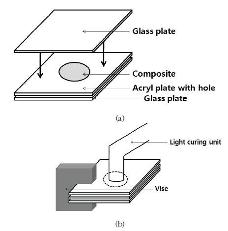
전치부 4급 복합레진 직접 수복을 모사하기 위하여 4 m 두께를 가지는 디스크 형태의 시편(직경 15 mm)을 제작하였다(Figure 1a).

S chematic d iagram o f specimen p reparation. (a) Composite was filled in the hole of an acryl plate above a glass plate and covered with another glass plate. (b) The glass and acryl plates were pressed with a vise and light cured.
미세혼합형복합레진(Z250)과 나노복합레진(Z350XT)의 3가지 shade 사이의 색차 및 각각의 투명도를 비교, 분석하기 위해 Z250/A2, Z350XT/A2E, Z350XT/A2B, Z350XT/A2D로 4개의 group (Group 1-4)에 대하여 각각 3개씩 총 12개의 시편을 제작하였다(Table 2). 나노복합레진 단일층과 적층 시의 색차 및 투명도를 비교하기 위해 Z350XT/A2E 1 mm + Z350XT/A3D 3 mm, Z350XT/A2E 2 mm + Z350XT/A3D 2 mm, 그리고 Z350XT/A2E 3 mm + Z350XT/A3D 1 mm 등 3개의 group (Group 5-7)에 대하여 각각 3개씩 총 9개의 시편을 제작하였다(Table 2).
유리판 위에 15 mm 직경의 구멍을 형성한 투명 아크릴판(각 group에 따라 1, 2, 3, 4 mm 두께)을 올리고 복합레진을 bulk 충전하고 다른 유리판을 상부에 위치시켜 vise를 이용하여 압착한 후 여분의 복합레진을 제거하고 광중합(두께≤2 mm enamel & body shade: 20초, 2 mm<두께≤4 mm enamel & body, 두께≤2 mm dentin shade: 40초, 2 mm<두께≤4 mm dentin shade: 80초) 하였다(Figure 1b). 두 가지 색상으로 적층하는 시편의 경우 dentin shade를 먼저 충전하여 광중합한 후 그 위에 enamel shade를 적층하였고 두 층 사이에 어떠한 처리도 하지 않았다.
제작한 시편은 spectrophotometer (CM3500d, Konica Minolta, Tokyo, Japan)를 이용하여 표준광원 D65하에서 reflectance 측정 모드로 배경이 없는 상태로 L*, a*, b*값을 측정하였다. 분광광도계의 target mask의 측정 부위 직경은 11 mm, 시편의 직경은 15 mm로 시편이 target mask 측정 부위를 완전히 덮도록 시편을 위치시켰다(Figure 2).
측정한 L*, a*, b*값을에 적용하여 단일층으로 적용한 Z250/A2 (Group 1)과 Z350XT의 3가지 shade A2E, A2B, A2D (Group 2, 3, 4) 사이의 색차 및 Z350XT/A2B (Group 3)를 단일층으로 적용한 경우와 Z350XT/A2E와 Z350XT/A3D를 적층하여 적용한 경우(Group 5, 6, 7) 사이의 색차(ΔE)를 구하였다.
TP (Translucency Parameter)는 같은 시편에 대하여 2 mm 두께의 검은색 아크릴판과 2 mm 두께의 흰색 아크릴판의 서로 다른 두 배경 조건에서 L*, a*, b*값을 측정한 후 (W: 흰색 배경, B: 검은색 배경) 공식에 적용하여 구했다.
3. 통계분석
통계분석은 실험군 사이의 L*, a*, b*값 비교, Z250과 Z350XT의 3가지 shade 사이의 색차 및 단일층 Z350XT와 적층 Z350XT 사이의 색차 비교, Z250, Z350XT 3가지 shade 각각의 투명도 및 Z350XT를 적층한 경우들 사이의 투명도 비교를 위해 일원배치 분산분석(one-way ANOVA)과 post-hoc Tukey’s test를 이용해 분석하였다. 모든 통계 분석은 SPSS software (version 21.0)을 이용하여 이루어졌으며, 검정을 위한 유의수준은 5%로 설정하였다.
Ⅲ. 결 과
단일층으로 적용한 Z250/A2의 L*값은 Z350XT/A2E, Z350XT/A2B, Z350XT/A2D 보다 작았고, a*값은 Z350XT/ A2E와 Z350XT/A2B 사이, 그리고 b*값은 Z350XT/A2B와 Z350XT/A2D 사이의 값을 보였다. Z350XT/A2 단일층 적용에서 L*, b*값은 enamel, body, dentin 순으로 증가하였다(p<0.05). 두 가지 색상으로 적층한 경우는 Z350XT/A2E의 두께가 증가할수록 L*, a*, b*값이 감소하였다(p<0.05, Table 3).
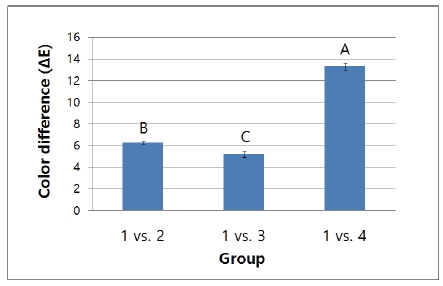
Z250과 Z350XT의 3가지 shade 사이의 색차(ΔE)를 비교한 경우 Z250/A2 (Group 1)과 Z350XT/A2E, Z350XT/A2B, Z350XT/A2D (Group 2, 3, 4) 사이의 색차는 순차적으로 6.26, 5.20, 13.36 이었다(Figure. 3).

Color difference (ΔE) between Z250/A2 (Group 1) and Z350XT/A2E, Z350XT/A2B, Z350XT/A2D (Group 2, 3, 4).
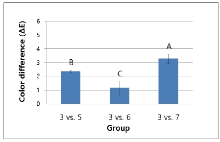
Z350XT/A2B를 단일층으로 적용한 경우(Group 3)와 Z350XT/A2E와 Z350XT/A3D를 두 층으로 적층한 경우 사이의 색차(ΔE)는 A2E 2 mm + A3D 2 mm로 적층했을 때(Group 6)가 1.19로 통계적으로 유의하게 가장 작았다(p<0.05, Figure 4).

Color difference between bulk-filled (Z350XT/A2B) and two-layered (Z350XT/A2E and Z350XT/A3D) composites.
Trasnlucency parameter (TP)는 단일층으로 적용한 Z250/A2 및 Z350XT/A2E, Z350XT/A2B, Z350XT/A2D 상호간에 통계적으로 유의한 차이를 나타내었다(p<0.05). 적층한 경우 A2E 두께가 증가할수록 유의하게 TP가 증가하였다(p<0.05, Figure 5). TP를 구하기 위해 사용한 흰색 배경과 검은색 배경의 L*, a*, b*값은 Table 4와 같다.
Ⅳ. 고 찰
본 연구에서는 심미가 중시되는 4급 와동의 복합레진 직접수복 시 여러 색상으로 적층하는 방법의 타당성을 알아보기 위해 같은 색상의 미세혼합형복합레진(Z250)과 나노복합레진(Z350XT)의 3가지 shade 사이의 색차 및 Z350XT를 단일층으로 적용한 경우와 두 가지 색상으로 적층한 경우 사이의 색차, 단일층으로 적용한 Z250, Z350XT enamel, body, dentin 각각의 투명도 및 Z350XT를 적층한 경우의 투명도를 분광광도계를 이용하여 측정한 L*, a*, b*을 통해 비교, 분석하였다.
한국인 상악중절치의 치관부 두께는 남성 7.10 mm, 여성 6.78 mm로, 치경부 두께는 남성 6.62 mm, 여성 6.43 mm로 보고되었으며(Paik과 Kim, 1988), 한국인 상악측절치의 치관부 두께는 남성 6.5 mm, 여성 6.2 mm로, 치경부 두께는 남성 6.1 mm, 여성 5.9 mm로 보고되어(Paik과 Kim 1989), 절단면을 포함한 4급 와동 수복을 모사하기 위해 시편의 두께는 4 mm로 설정하였다.
상악중절치의 측정위치에 따른 색과 투명도를 분광광도계를 이용하여 분석한 연구에서 a*, b*값은 절단부에서 치경부로 갈수록 값이 증가하였으며 투명도는 감소하였다(Haseqawa 등, 2000). 또한 복합레진의 shade 숫자가 증가할수록 a*, b*값이 증가하였다(Park과 Lee, 2007). 이에 따라 본 연구에서는 치아 중앙부 4급 와동 수복에 대하여 진료실에서 가장 많이 이용되는 A2 shade를 기준 색상으로 하여 실험군을 설정하였다.
색차값(ΔE)>2 이면 모든 관찰자에서 색차를 정확히 인지하고, 1<ΔE<2 이면 관찰자의 부정확한 판단 비율이 높아지며 ΔE1<1 일 경우 거의 인지할 수 없다(Seghi 등, 1989). 또한 50%의 관찰자가 수용할 수 있는(acceptable) 색차의 상한선은 2.72(Ragain과 Johnston, 2000), 또는 3.3(Ruyter 등, 1987)으로 제시된 바 있다. Z250/A2와 Z350XT /A2E, Z350XT/ A2B, Z350XT/A2D의 3가지 shade들의 색차를 비교하였을 때, 모두 3.3보다 큰 값을 나타내어 관찰자에 의해 수용될 수 없는 수준이었고, 각각의 투명도를 비교한 결과 모두 통계적으로 유의한 차이를 나타내어 Z250을 대체하여 Z350XT를 임상에서 사용할 때 색차 및 투명도 차이에 유의해야 할 것이다.
제조사는 4급 와동을 Z350XT 단일 색상으로 수복할 경우 Vita 색조가이드에 대응되는 body shade를 선택하도록 제시하고 있으며, 여러 색상으로 적층하여 수복하는 경우에는 body shade와 동일한 shade의 enamel shade와 한 단계 큰 숫자의 dentin shade를 겹쳐서 충전할 것을 제시하고 있다. 법랑질 본연의 투명도와 두께로 인하여 내부 상아질의 채도가 감소된다는 연구결과가 있으며(Dietschi, 2001), 본 연구결과에 의하면 enamel shade는 body와 dentin shade에 비하여 L*, a*, b*값이 작기 때문에 한 단계 큰 숫자의 dentin shade를 선택하도록 제시한 것은 타당하다고 볼 수 있다. 또한 4급 와동의 경우 검은색에 가까운 구강 내부의 배경색을 차단해야 하며 와동이 깊기 때문에 body shade 보다는 불투명 성향이 더 큰 dentin shade를 선택하도록 제시한 것으로 생각된다.
제조사의 지시에 따라 두 층으로 적층할 경우 enamel shade와 dentin shade 두께의 적절한 비율을 알아보기 위해 Z350XT/A2B를 단일층으로 적용한 경우와 Z350XT/A2E와 Z350XT/A3D를 적층하여 적용한 경우 사이의 색차를 비교하였는 바, A2E 2 mm + A3D 2 mm로 적층했을 때가 통계적으로 유의하게 가장 작은 값을 나타내었다. 심미수복재료의 색과 투명도는 복합레진 기질의 종류와 충전재의 조성과 양 뿐만 아니라 미량의 색소첨가 및 기타 화학성분에 의해서도 변화 할 수 있다(Johnston과 Reisbick, 1997). 같은 복합레진 기질과 충전재를 가지는 Z350XT enamel, body, dentin 각각에 포함된 색소의 양과 종류에 따라 서로 다른 색을 나타낼 수 있으며, enamel shade의 두께 증가에 따른 투명도 증가도 적층한 복합레진의 색을 변화시킬 수 있다. 이러한 요인들이 복합적으로 작용하여 A2E 2 mm + A3D 2 mm로 적층했을 때가 A2B와 비교 시 가장 작은 색차를 나타냈을 것이다.
동일한 색상의 복합레진을 적층 충전하였을 경우 bulk 충전과 비교하여 직선 투과와 TP는 감소하고 확산 투과는 증가하였는데(Horie 등, 2012), 이번 연구에서 enamel shade와 dentin shade를 적층한 경우 enamel shade의 두께가 증가할수록 투명도가 증가한 것은 적층에 의해 나타나는 복합레진 층간 부위의 resin-rich zone에서 일어나는 광투과 특성의 변화 보다는 enamel shade 자체가 가지는 높은 투명도가 더 큰 영향을 미친 결과로 생각된다.
본 연구에서는 11 mm 직경의 원형 구경(aperture)의 분광광도계를 사용하였다. 구경 크기가 커지면 TP 값이 증가한다고 하였는데(Yu 등, 2009), 이는 실제 치아 크기보다 다소 큰 구경이므로 실제 임상에서 치아 와동의 충전에 사용되는 크기의 복합레진의 TP 값과는 차이가 있을 수 있다. 또한 A2 shade를 기준 색상으로 하여 얻어진 결과로 추후 더욱 다양한 shade를 이용한 실험군을 설정한 연구가 필요할 것이다.
본 연구에서 미세혼합형복합레진(Z250)과 나노복합레진(Z350XT)의 enamel, body, dentin shade 사이의 색차를 비교했을 때 수용되기 어려울 정도의 색차(ΔE>3.3)를, 나노복합레진을 단일층으로 적용한 경우와 여러 색상으로 적층한 경우들 사이에서는 수용될 수 있는 색차(ΔE<3.3)를 나타내어 첫 번째와 두 번째 귀무가설은 기각되었다. 또한 단일층으로 적용한 미세혼합형복합레진, 나노복합레진 enamel, body, dentin 각각의 투명도 및 나노복합레진을 여러 색상으로 적층한 경우의 투명도 사이에는 유의한 차이를 보여서 세 번째 귀무가설도 기각되었다.
Ⅴ. 결 론
실험실 연구의 한계 내에서, 미세혼합형복합레진(Z250)과 나노복합레진(Z350XT)의 3가지 shade (enamel, body, dentin) 사이의 색차를 비교했을 때 수용되기 어려울 정도의 색차(ΔE>3.3)를 가지며 각각의 투명도가 유의한 차이를 보이므로 임상 적용 시 나노복합레진 각 shade들을 구분하여 사용해야 할 필요성을 알 수 있었다.
Z350XT/A2B를 단일층으로 적용한 경우와 Z350XT/A2E와 Z350XT/A3D를 적층한 경우들 사이의 색차는 A2E 2 mm + A3D 2 mm 시편이 통계적으로 유의하게 가장 낮은 색차를 나타내었고, 적층한 시편들 사이의 투명도 차이는 enamel shade 두께가 두꺼워질수록 투명도가 증가하였다.
결론적으로 Z350XT를 이용하여 A2 shade 4 mm 두께의 4급 와동을 두 층으로 적층하여 수복 시에는 A2E 2 mm + A3D 2 mm 적용이 추천된다.
Acknowledgments
이 연구는 한국연구재단의 기초과학연구프로그램에 의한 교육부의 지원(No. 2016R1D1A1B03931827)에 의해 이루어졌음.
References
-
Azzopardi, N, Moharamzadeh, K, Wood, DJ, Martin, N, van Noort, R, (2009), Effect of resin matrix composition on the translucency of experimental dental composite resins, Dent Mater, 25(12), p1564-8.
[https://doi.org/10.1016/j.dental.2009.07.011]

- Darabi, F, Radafshar, G, Tavangar, M, Davaloo, R, Khosravian, A, Mirfarhadi, N, (2014), Translucency and masking ability of various composite resins at different thicknesses, J Dent Shiraz Univ Med Sci, 15(3), p117-22.
- Dietschi, D, (2001), Layering concepts in anterior composite restorations, J Adhes Dent 3, (1), p71-80.
- Dietschi, D, Ardu, S, Krejci, I, (2006), A new shading concept based on natural tooth color applied to direct composite restorations, Quintessence Int, 37(2), p91-102.
-
Dietschi, D, (2008), Optimising aesthetics and facilitating clinical application of free-hand bonding using the ‘natural layering concept’, Br Dent J, 204(4), p181-5.
[https://doi.org/10.1038/bdj.2008.100]

-
Dos Santos, GB, Alto, RV, Filho, HR, Da Silva, EM, Fellows, CE, (2008), Light transmission on dental resin composites, Dent Mater, 24(5), p571-6.
[https://doi.org/10.1016/j.dental.2007.06.015]

-
Goodkind, RJ, and Schwabacher, WB, (1987), Use of a fiber-optic colorimeter for in vivo color measurements of 2830 anterior teeth, J Prosthet Dent, 58(5), p535-42.
[https://doi.org/10.1016/0022-3913(87)90380-5]

-
Hajira, NS, Mehta, D, Ashwini, P, Meena, N, Usha, HL, (2015), Influence of Different Enamel Shades and Thickness on Chroma and Value of Dentin Vita Shade: An in vitro Comparative Assessment Study, J Contemp Dent Pract, 16(4), p304-9.
[https://doi.org/10.5005/jp-journals-10024-1680]

-
Hasegawa, A, Ikeda, I, Kawaguchi, S, (2000), Color and translucency of in vivo natural central incisors, J Prosthet Dent, 83, p418-23.
[https://doi.org/10.1016/S0022-3913(00)70036-9]

-
Horie, K, Nakajima, M, Hosaka, K, Kainose, K, Tanaka, A, Foxton, RM, Tagami, J, (2012), Influences of composite-composite join on light transmission characteristics of layered resin composites, Dent Mater, 28(2), p204-11.
[https://doi.org/10.1016/j.dental.2011.10.006]

-
Ikeda, T, Sidhu, SK, Omata, Y, Fujita, M, Sano, H, (2005), Colour and translucency of opaque-shades and body-shades of resin composites, Eur J Oral Sci, 113(2), p170-3.
[https://doi.org/10.1111/j.1600-0722.2005.00205.x]

-
Johnston, WM, Reisbick, MH, (1997), Color and translucency changes during and after curing of esthetic restorative materials, Dent Mater, 13, p89-97.
[https://doi.org/10.1016/S0109-5641(97)80017-6]

-
Jung, JE, Jung, KH, Son, SA, Hur, B, Kwon, YH, Park, JK, (2014), Comparison of the color of composite resins with different thickness, Korean J Dent Mater, 41(2), p85-93.
[https://doi.org/10.14815/kjdm.2014.41.2.85]

-
Kamishima, N, Ikeda, T, Sano, H, (2006), Effect of enamel shades on color of layered resin composites, Dent Mater, 25(1), p26-31.
[https://doi.org/10.4012/dmj.25.26]

-
Khashayar, G, Dozic, A, Kleverlaan, CJ, Feilzer, AJ, Roeters, J, (2014), The influence of varying layer thicknesses on the color predictability of two different composite layering concepts, Dent Mater, 30(5), p493-8.
[https://doi.org/10.1016/j.dental.2014.02.002]

-
Kim, BJ, & Lee, YK, (2009), Influence of the shade designation on the color difference between the same shade-designated resin composites by the brand, Dent Mater, 25(9), p1148-54.
[https://doi.org/10.1016/j.dental.2009.04.001]

-
Kim, SJ, Son, HH, Cho, BH, Lee, IB, Um, CM, (2009), Translucency and masking ability of various opaque-shade composite resins, J Dent, 37(2), p102-7.
[https://doi.org/10.1016/j.jdent.2008.10.002]

-
Lee, YK, (2008), Influence of filler on the difference between the transmitted and reflected colors of experimental resin composites, Dent Mater, 24(9), p1243-7.
[https://doi.org/10.1016/j.dental.2008.01.014]

-
Lee, YK, Yu, B, Lee, SH, Cho, MS, Lee, CY, Lim, HN, (2010), Shade compatibility of esthetic restorative materials-A review, Dent Mater, 26(12), p1119-26.
[https://doi.org/10.1016/j.dental.2010.08.004]

-
Lee, YK, (2015), Translucency of human teeth and dental restorative materials and its clinical relevance, J Biomed Opt, 20(4), p045002-1-8.
[https://doi.org/10.1117/1.JBO.20.4.045002]

- Lim, YK, Lee, YK, Lim, BS, Rhee, SH, Yang, HC, (2008), Influence of filler distribution on the color parameters of experimental resin composites, Dent Mater, 24(1), p67-73.2.
- Magne, P, and So, WS, (2008), Optical integration of incisoproximal restorations using the natural layering concept, Quintessence Int, 39(8), p633-43.
-
Naeimi Akbar, H, Moharamzadeh, K, Wood, DJ, Van Noort, R, (2012), Relationship between color and translucency of multishaded dental composite resins, Int J Dent, 2012, p708032-1-5.
[https://doi.org/10.1155/2012/708032]

-
O’Brien, WJ, (1997), Color distribution of three regions of extracted human teeth, Dent Mater, 13(3), p179-85.
[https://doi.org/10.1016/S0109-5641(97)80121-2]

- Paik, KS, and Kim, MK, (1988), Statistical Study on Morphology and Size of the maxillary Central Incisor in Korea Adult, The Korean Journal of Oral Anatomy, 11(1), p37-42.
- Paik, KS, and Kim, MK, (1989), A Study on Morphology and Size of the Maxillary Lateral Incisor in Korean Adult, The Korean Journal of Physical Anthropology, 2(2), p95-112.
- Park, DR, Kim, AJ, Bae, JM, Oh, SH, (2015), The fluorescence Evaluation of composite resin by digital imaging device and monochrome color analysis software, Korean J Dent Mater, 42(2), p73-82.
-
Park, JW, (2011), Understanding of the color in composite resin, J Kor Acad Cons Dent, 36(4), p271-9.
[https://doi.org/10.5395/JKACD.2011.36.4.271]

- Park, SK, and Lee, YK, (2007), Shade distribution of commercial resin composites and color difference with shade guide tabs, Am J Dent, 20(5), p335-9.
-
Ragain, JC, and Johnston, WM, (2000), Color acceptance of direct dental restorative materials by human observers, Color Res Appl, 25, p278-85.
[https://doi.org/10.1002/1520-6378(200008)25:4<278::AID-COL8>3.0.CO;2-F]

-
Ruyter, IE, Nilner, K, Moller, B, (1987), Color stability of dental composite resin materials for crown and bridge veneers, Dent Mater, 3, p246-51.
[https://doi.org/10.1016/S0109-5641(87)80081-7]

-
Seghi, RR, Hewlett, ER, Kim, J, (1989), Visual and instrumental colorimetric assessments of small color differences on translucent dental porcelain, J Dent Res, 68, p1760-4.
[https://doi.org/10.1177/00220345890680120801]

-
Ten Bosch, JJ, and Coops, JC, (1995), Tooth color and reflectance as related to light scattering and enamel hardness, J Dent Res, 74, p374-80.
[https://doi.org/10.1177/00220345950740011401]

-
Villarroel, M, (2011), Direct esthetic restorations based on translucency and opacity of composite resins, J Esthet Restor Dent, 23(2), p73-87.
[https://doi.org/10.1111/j.1708-8240.2010.00392.x]

-
Yu, B, Ahn, JS, Lee, YK, (2009), Measurement of translucency of tooth enamel and dentin, Acta Odontol Scand, 67(1), p57-64.
[https://doi.org/10.1080/00016350802577818]


