
Effect of additional firing and surface roughness on the mechanical behaviors of lithium disilicate glass ceramics

Abstract
This study aimed to evaluate the combined effects of surface roughness and additional firing cycles on the flexural strength of lithium disilicate (LS2) glass-ceramics using clinically relevant polishing protocols.One hundred and five bar-shaped LS2 specimens (3.0×4.0×30.0 mm) were divided into seven groups: one control group [EXF] and six experimental groups categorized by three surface roughness levels—fine [FI], medium [ME], and coarse [CO] with or without an additional firing cycle (840℃ for 5 minutes). Surface roughness was controlled using silicon carbide papers. All specimens underwent thermocycling (10,000 cycles, 5℃/55℃) before testing. Flexural strength was measured via a three-point bending test. Surface topography and phase transitions were analyzed using atomic force microscopy (AFM), X-ray diffraction (XRD), and scanning electron microscopy (SEM).Flexural strength was significantly higher in the EXF, FI, and FI_R groups compared to the ME, ME_R, CO, and CO_R groups (p<0.001). While additional firing alone did not significantly affect flexural strength (p>0.05), a significant interaction between surface roughness and firing was observed (p<0.01). SEM analysis revealed crack arrest lines and smoother fracture features in the lower-roughness groups. XRD patterns indicated an increased SiO2 content in specimens subjected to additional firing. Surface roughness significantly affects the flexural strength of LS2 ceramics. Additional firing modulates this effect depending on surface condition. Clinicians should prioritize smooth surface finishes and carefully consider post-adjustment firing protocols.
초록
본 연구의 목적은 임상적으로 유효한 연마 프로토콜을 적용하여 표면 거칠기와 추가 소성 사이클이 리튬 디실리케이트(LS2) 글래스 세라믹의 굽힘 강도에 미치는 복합적인 영향을 평가하는 것이다. 105개의 바(bar) 형태 LS2 시편(3.0×4.0×30.0 mm)을 제작하여 대조군(EXF)과 표면 거칠기 수준(Fine [FI], Medium [ME], Coarse [CO]) 및 추가 소성(840℃ for 5 minutes) 여부에 따른 6개의 실험군 등 총 7개 그룹(n=15)으로 분류하였다. 표면 거칠기는 실리콘 카바이드 페이퍼를 사용하여 조절하였다. 모든 시편은 강도 시험 전 열순환 thermocycling (10,000 cycles, 5℃/55℃) 과정을 거쳤다. 굽힘 강도는 3점 굽힘 시험으로 측정하였으며, 원자간력 현미경(AFM), X선 회절 분석(XRD), 주사전자현미경(SEM)을 이용하여 표면 지형 및 상 변화를 분석하였다. 굽힘 강도는 EXF, FI, FI_R 그룹이 ME, ME_R, CO, CO_R 그룹에 비해 유의하게 높게 나타났다(p<0.001). 추가 소성 단독으로는 굽힘 강도에 유의한 영향을 미치지 않았으나(p>0.05), 표면 거칠기와 소성 사이에는 유의한 상호작용이 관찰되었다(p<0.01). SEM 분석 결과, 낮은 거칠기를 가진 그룹에서 균열 정지선(crack arrest lines)과 더 매끄러운 파절 특성이 관찰되었다. XRD 패턴 분석에서는 추가 소성을 거친 시편에서 SiO2 함량이 증가하는 경향을 보였다. 표면 거칠기는 리튬 디실리케이트 세라믹의 굽힘 강도에 결정적인 영향을 미친다. 추가 소성은 표면 조건에 따라 이러한 영향을 조절하는 역할을 한다. 본 연구의 결과는 임상적으로 리튬 디실리케이트 수복물의 조정 후 강도 회복을 도모할 때, 추가 소성처리에 의존하기보다는 철저하고 정밀한 단계별 기계적 연마 프로토콜을 수행하는 것이 구조적 안정성과 장기적인 임상 성공을 확보하는 데 훨씬 더 결정적이고 효과적인 방법임을 시사한다.
Keywords:
Lithium disilicate, Surface roughness, Flexural strength, Additional firing, Weibull modulus키워드:
리튬 디실리케이트, 표면 거칠기, 굽힘 강도, 추가 소성, 와이블 계수Introduction
Lithium disilicate (LS2) glass ceramics are widely used in fixed prosthodontics due to their superior optical properties and enhanced mechanical performance compared to conventional feldspathic porcelains (1, 2). Although LS2 does not exhibit transformation toughening like zirconia, its relatively high fracture resistance is primarily attributed to residual compressive stresses formed during sintering due to mismatched thermal expansion between the LS2 crystals and the surrounding glass matrix (1). Additionally, the elongated, interlocking crystal morphology contributes to crack deflection and bridging, further enhancing fracture resistance (3-6).
Optimal mechanical properties are typically achieved when the LS2 crystal phase presents a balanced combination of crystal size and aspect ratio, a condition influenced by thermal treatment (7). However, commercially available systems such as IPS e.max (1–1.5 µm) and others with smaller crystals (0.2–0.5 µm) have shown widely variable flexural strength, ranging from 360 to 700 MPa (6, 8). This variability suggests that surface conditions, in addition to microstructure, play a critical role in determining mechanical behavior.
Surface flaws introduced during clinical procedures such as grinding, contouring, and occlusal adjustments act as stress concentrators that compromise structural integrity (9, 10). Chairside polishing is commonly used to smooth these surfaces and restore strength (11). More recently, additional firing after polishing has been proposed as a method to enhance surface quality by inducing partial viscous flow in the glassy phase, potentially sealing microdefects and mitigating crack initiation (12, 13). For instance, Alves et al. reported improvements in surface smoothness and flexural strength following heat treatment (12), while Lu et al. demonstrated healing of surface microcracks with controlled firing (13, 14).
Despite these promising results, few studies have explored the combined effects of clinically relevant polishing protocols and short-duration additional firing on the mechanical behavior of LS2 ceramics. Moreover, prior research has often focused on single-material systems, overlooking the variability in chemical composition and microstructure among commercially available products (8, 10-12). Furthermore, while some investigations have shown that fine polishing improves strength, others have reported inconsistent correlations between surface roughness and flexural strength, likely due to differences in subsurface damage, crystal orientation, or phase composition (11, 15, 16).
Yahyazadehfar et al. emphasized that surface quality alone does not reliably predict ceramic strength due to the complex interplay of microcracks and flaw distributions (15). Similarly, Deville et al. highlighted the influence of residual stresses and surface finishing on the mechanical performance and aging resistance of ceramics (16). These findings underscore the multifactorial nature of surface integrity and its interaction with both intrinsic and extrinsic material properties.
Therefore, this in vitro study aimed to evaluate the effects of surface roughness and additional firing on the flexural strength of LS2 glass ceramics under simulated clinical conditions. The first null hypothesis was that surface roughness would have no effect on flexural strength. The second hypothesis was that additional firing would exert no effect, regardless of surface condition.
Materials and Methods
1. Specimen Preparation
This in vitro study evaluated the effects of clinically relevant surface roughness and additional short-duration firing on the flexural strength of lithium disilicate (LS2) glass-ceramics. A total of 105 bar-shaped specimens (3.0×4.0×30.0 mm) were fabricated from LS2 blocks (Amber Mill, C40/A2; HASSBIO, Gangneung, Korea). All specimens were chamfered at the edges (0.1 mm at 45°) in accordance with ISO 6872:2015 guidelines. The specimens were sectioned using a low-speed saw (Samsung Clover, Yangju, Korea) and sequentially polished with abrasive grains (6 µm, 3 µm, and 1 µm) using specialized polishing units (HRG-150, AM Technology, Asan, Korea; SPL-15 Grind X, OKAMOTO, Saitama, Japan). Crystallization was conducted in a ceramic furnace (Austromat 624i, Dekema, Freilassing, Germany) following the manufacturer’s instructions.
2. Experimental Grouping and Surface Treatment
Specimens were randomly assigned to seven groups (n=15 per group): one control group [EXF] and six experimental groups. The EXF group featured a highly polished surface (Ra 0.10-0.30 μm) to serve as a reference for maximum flexural strength. While the EXF group represented the ideal baseline condition with minimal surface flaws prepared by standard laboratory polishing protocols, the experimental fine group [FI] was intentionally ground and subsequently restored using clinically relevant chairside fine polishing protocols to simulate intraoral adjustments. Three levels of clinically relevant surface roughness were established: fine [FI: Ra 0.10–0.30 µm], medium [ME: Ra 0.80–1.60 µm], and coarse [CO: Ra 1.80–2.60 µm] using silicon carbide papers (CC261, Deerfos, Seoul, Korea). Each roughness category was further divided based on whether an additional firing cycle was performed (840℃ for 5 minutes), designated as FI_R, ME_R, and CO_R. Surface roughness was measured using a contact profilometer (TR200, TIME High Technology Ltd., Beijing, China), with five measurements averaged per specimen.
3. Aging and Mechanical Testing
All specimens were stored in distilled water at 37℃ for 24 hours, followed by thermocycling for 10,000 cycles (5℃-55℃, 30-second dwell time, 2-second transfer) using a thermal cycling unit (R&B Inc., Daejeon, Korea). Flexural strength was evaluated using a universal testing machine (Instron 5982, Instron Corp., Norwood, MA, USA) in a three-point bending configuration with a 30 mm span and 1 mm/min crosshead speed. The Weibull modulus was calculated according to ISO 6872:2015.
4. Microstructural and Phase Analysis
Fractographic analysis was performed using scanning electron microscopy (SEM; Quanta FEG 250, FEI, Hillsboro, USA) at 50×magnification. High-resolution surface imaging was conducted via field-emission SEM (FE-SEM; SU5000, Hitachi, Tokyo, Japan) at 20,000× magnification. Phase composition was analyzed using X-ray diffraction (XRD; Empyrean, Malvern Panalytical, Malvern, UK) with Cu-Kα radiation source (45 kV, 40 mA), scanning from 2θ= 10°-80° (step size: 0.131303°, dwell time: 88.995 seconds). Quantitative analysis was performed via Rietveld refinement (HighScore Plus software). Surface topography was further characterized using atomic force microscopy (AFM; ezAFM, NanoMagnetics Instruments, Oxford, UK) over a 60µm×60 µm area.
5. Statistical Analysis
Data were analyzed using IBM SPSS Statistics 25.0 (Armonk, NY, USA). Normality and homogeneity of variances were verified using the Shapiro-Wilk and Levene’s tests (p>0.05). One-way ANOVA followed by Tukey's HSD post hoc test was performed to determine significant differences among groups (α=0.05). A two-way ANOVA analyzed the interaction between surface roughness and additional firing.
Results
1. Flexural strength and group comparisons
Mean flexural strength varied significantly according to surface roughness (Table 1, Figure 1). The EXF, FI, and FI_R groups demonstrated the highest mean flexural strength values (mean ± SD), whereas the ME, ME_R, CO, and CO_R groups showed significantly lower values (p< 0.001) (Figure 1). Among all groups, the EXF group exhibited the greatest strength, underscoring the favorable effect of minimal surface irregularity. One-way ANOVA confirmed statistically significant differences among the roughness groups (p< 0.001).

Two-way ANOVA result for flexural strength of experimental groups
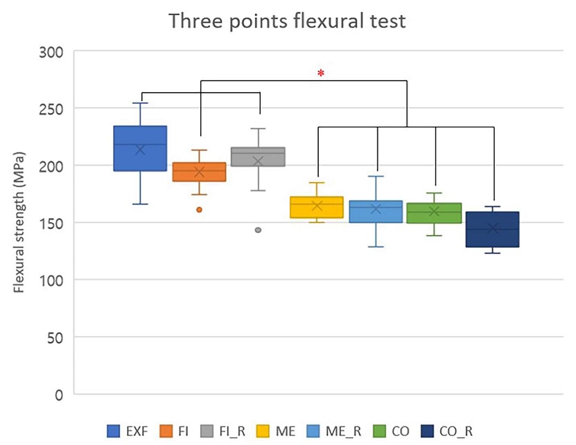
Box plot of comparing the flexural strength of each group.
2. Interaction effects of surface roughness and additional firing
Two-way ANOVA revealed a significant main effect of surface roughness (p< 0.001), while the main effect of additional firing alone was not statistically significant (p> 0.05) (Table 1). However, a significant interaction between surface roughness and additional firing was observed (p< 0.01), indicating that the effect of firing was modulated by surface condition (Table 1). In particular, the CO_R group showed a more pronounced reduction in flexural strength compared to the ME_R and FI_R groups (Table 1, Figure 1). The equal group sizes (n=15 per subgroup) ensured a balanced two-way ANOVA design.
To further interpret the interaction, simple effects analysis was conducted. When surface roughness was held constant, additional firing did not induce statistically significant changes in flexural strength within the FI and ME groups (p> 0.05), although a slight decrease was noted in the CO group. In contrast, across all firing conditions, flexural strength significantly decreased as surface roughness increased (FI > ME > CO; p<0.001) (Figure 1).
3. Structural reliability and surface roughness characteristics
Weibull analysis showed the highest modulus in the ME group and the lowest in the FI_R group (Figure 2). Non-fired groups demonstrated greater reliability (modulus range: 16.02-19.44), whereas fired groups exhibited reduced Weibull moduli (10.09-12.14), indicating broader strength distributions following firing (Figure 2).
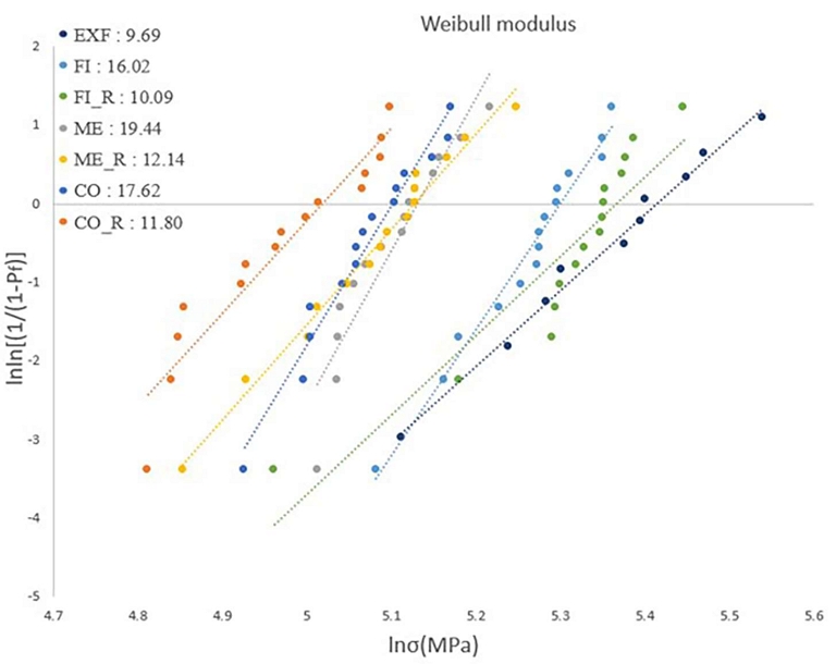
Weibull plot of each group.
Surface roughness measurements before and after firing revealed no significant differences within the FI_R, ME_R, and CO_R groups (p> 0.05), suggesting that the additional firing protocol did not substantially alter surface texture. This statistical lack of difference was visually and topographically confirmed via atomic force microscopy (AFM) analysis, which demonstrated highly consistent surface profiles and peak-to-valley heights between the respective non-fired and fired subgroups (Figure 6). However, overall comparisons indicated significantly lower Ra values in the FI and FI_R groups compared to the others (p< 0.001) (Figure 6).
4. Microstructural, fractographic, and phase analyses
SEM analysis of fractured surfaces revealed crack arrest lines in the FI and FI_R groups only, indicating improved crack resistance associated with smoother surfaces (Figure 3). In contrast, continuous crack propagation was observed in the remaining groups (Figure 3). High-magnification SEM imaging revealed an increased number of pores in the glass phase among specimens subjected to additional firing, particularly in the ME_R and CO_R groups (Figure 4).
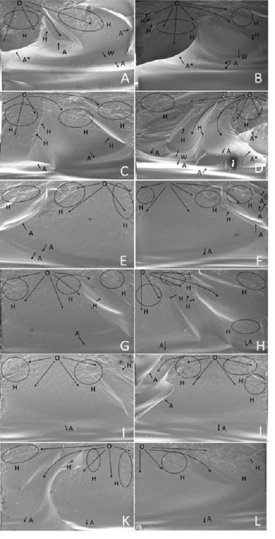
Scanning electron microscope images of fractured surfaces. (magnification x50) A, B: FI group, C, D: FI_R group, E, F: ME group, G, H: ME_R group, I, J: CO group, K, L: CO_R group, dotted line: crack propagation line, O: origin of fracture, H: hackles, W: Wallner line, A: arrest line, A*: arrest line that prevented the crack propagation from penetrating the opposite surface.
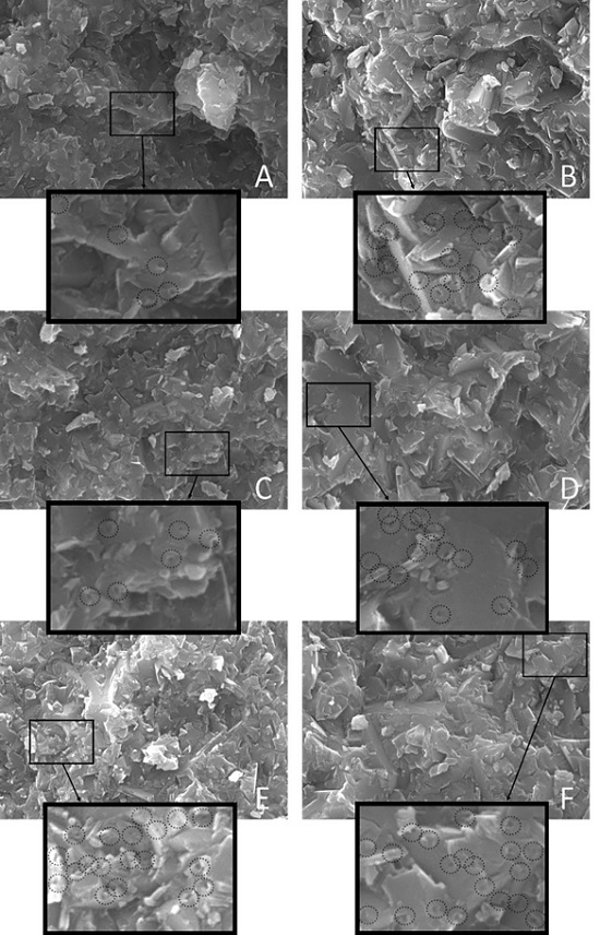
Scanning electron microscope images (magnification x20,000). Additional firing groups (B, D, F) tend to exhibit a consistent presence of numerous pores in the glass matrix. A: FI, B: FI_R, C: ME, D: ME_R, E: CO, F: CO_R.
XRD analysis identified peaks corresponding to Li2Si2O5 and SiO2 in all groups (Figure 5). Notably, fired specimens exhibited increased SiO2 content, suggesting partial re-precipitation of the glassy phase during the firing cycle (Figure 5). AFM imaging showed progressively greater topographic irregularity with increasing surface roughness, with more pronounced peak-to-valley differences observed in the CO, ME, CO_R, and ME_R groups (Figure 6).
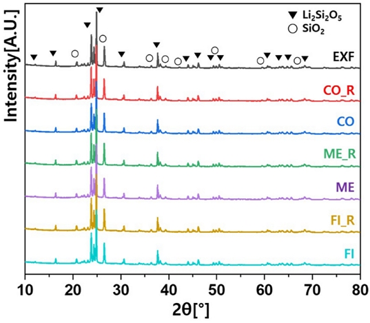
X-ray diffraction patterns of samples.
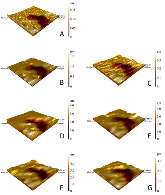
Atomic force microscopy (AFM) images of each group. The scale bars represent the peak-to-valley height of the surface roughness. Darker areas indicate relatively deep defects compared to the surrounding areas. A: EXF, B: FI, C: FI_R, D: ME, E: ME_R, F: CO, G: CO_R.
Taken together, these findings demonstrate that surface roughness is a primary determinant of flexural strength in LS2 ceramics. The effect of additional firing appears to be condition-dependent and may adversely affect strength under certain surface conditions.
Discussion
This study evaluated the combined effects of clinically relevant surface roughness and short-duration additional firing on the flexural strength of lithium disilicate (LS2) glass ceramics. Increased surface roughness significantly compromised strength, leading to the rejection of the first null hypothesis. In contrast, additional firing alone did not significantly alter strength values, supporting the second null hypothesis.
Atomic force microscopy (AFM) confirmed that groups with moderate to high roughness (ME, ME_R, CO, CO_R) exhibited deeper surface irregularities, correlating with significantly reduced flexural strength (p< 0.001).
Scanning electron microscopy (SEM) revealed crack arrest lines in the FI and FI_R groups, suggesting enhanced crack resistance in smoother surfaces. These findings align with previous studies demonstrating that surface flaws such as microcracks act as stress concentrators under tensile stress, thus reducing mechanical reliability (2-4, 11, 12, 16, 31).
Although fine polishing has been shown to improve flexural strength, other reports highlight variability in outcomes, even among similarly polished specimens (11, 16, 17) This inconsistency may stem from variations in subsurface damage or polishing-induced microstructural changes (16).
Two-way ANOVA in this study demonstrated a significant interaction between surface roughness and additional firing (p< 0.01), although neither factor showed a standalone effect. SEM at 20,000×magnification revealed elevated pore density in fired groups, especially ME_R and CO_R, which may explain the reduced Weibull modulus and greater strength variability (Figure 4).
Regarding the nature of these pores, the short-duration additional firing protocol (840℃ for 5 minutes) possessed insufficient thermal energy to alter the entire bulk internal structure of the LS2 glass-ceramics. Instead, the increased pore density is highly likely a subsurface phenomenon, wherein the softened glass matrix under thermal treatment flowed into and trapped micro-voids within the pre-existing subsurface damage layer induced by grinding. This localized structural alteration undermines the mechanical reliability of the material, as evidenced by the decreased Weibull modulus, without necessarily triggering a significant drop in the overall mean flexural strength of the bulk material. This pattern is consistent with previous literature linking thermally induced porosity to decreased mechanical reliability. This pattern is consistent with previous literature linking thermally induced porosity to decreased mechanical reliability (23).
The dual impact of additional firing—both as a surface healing mechanism and a potential source of internal defects—was further supported by increased SiO2 peaks in XRD and AFM-observed surface topography changes. These findings corroborate earlier reports that highlight the complexity of thermal processing in LS2 ceramics (20, 30).
Previous studies have yielded conflicting conclusions on the effects of repeated firing, with some showing strength reduction (22-24) and others reporting neutral or even beneficial effects (12, 14, 18, 25-27) These discrepancies may be attributed to differing firing parameters, ceramic compositions, and surface preparation techniques (19, 22-24).
The Ra values targeted in this study (0.10–0.30 µm, 0.80–1.60 µm, 1.80–2.60 µm) were successfully achieved and are consistent with clinically relevant polishing levels reported in the literature (11, 16, 28-30, 32) While minor variability may be due to material-specific or environmental factors (16, 28), the statistically significant differences among groups support the validity of the experimental design.
This study has several limitations. Only one commercial LS2 product was evaluated, which may limit generalizability (8). Furthermore, while silicon carbide papers allowed for precise roughness control, they do not fully replicate intraoral finishing conditions. Future studies should explore a broader range of LS2 materials, firing protocols, and clinically representative surface treatments. Additional investigations involving glaze applications or modified sintering cycles may also help determine the threshold at which firing transitions from beneficial to detrimental.
In conclusion, surface roughness is a critical determinant of LS2 ceramic strength. Although additional firing may facilitate surface healing, its overall mechanical benefit is contingent upon the existing surface condition and potential internal structural changes. Clinically, these results underscore the importance of meticulous polishing and a judicious approach to post-adjustment thermal treatments.
The clinical implications of the present study are as follows. First, since surface roughness is the primary determinant of the flexural strength of LS2 glass-ceramics, meticulous mechanical polishing is indispensable following any occlusal or contour adjustments. Achieving a high-gloss, smooth surface comparable to the EXF or FI groups in this study is essential to preserve the material’s inherent structural integrity. Second, the common clinical assumption that additional firing can “heal” surface micro- cracks or defects was not supported by our findings; instead, short-duration firing on moderately or highly roughened surfaces may lead to a reduction in mechanical reliability, as evidenced by the decreased Weibull modulus. Therefore, rather than relying on additional firing as a remedial measure for surface damage, clinicians should adhere to a strict protocol of sequential polishing to ensure the long-term durability and success of LS2 restorations in clinical practice.
Conclusion
Within the limitations of this study, the following conclusions were drawn:
- 1. Surface roughness is the dominant factor influencing the flexural strength of LS2 glass-ceramics, with smoother surfaces significantly enhancing strength and crack resistance.
- 2. Increased surface roughness compromises mechanical performance, and this negative effect is not mitigated by subsequent thermal treatments
- 3. Additional firing interacts significantly with surface condition; it may reduce the mechanical reliability (Weibull modulus) of restorations with moderate to high surface roughness.
- 4. Meticulous polishing remains clinically essential to ensure the structural durability of LS2 restorations, especially after occlusal adjustments, whereas the routine use of additional firing should be performed with caution.
clinical practice.
Acknowledgments
This work was supported by the 2024 Gangneung-Wonju National University Dental Hospital Scientific Research Grant (SR2401).
References
-
Zarone F, Di Mauro MI, Ausiello P, Ruggiero G, Sorrentino R. Current status on lithium disilicate and zirconia: a narrative review. BMC Oral Health. 2019;19:134.
[https://doi.org/10.1186/s12903-019-0838-x]

- Porto TS, Roperto RC, Akkus A, Akkus O, Teich S, Faddoul FF, et al. Effect of thermal cycling on fracture toughness of CAD/CAM materials. Am J Dent. 2018;31:205-10.
-
Hallmann L, Ulmer P, Kern M. Effect of microstructure on the mechanical properties of lithium disilicate glass ceramics. J Mech Behav Biomed Mater. 2018;82:355-70.
[https://doi.org/10.1016/j.jmbbm.2018.02.032]
-
Zhang Z, Guo J, Sun Y, Tian B, Zheng X, Zhou M, et al. Effects of crystal refining on wear behaviors and mechanical properties of lithium disilicate glass ceramics. J Mech Behav Biomed Mater. 2018;81:52-60.
[https://doi.org/10.1016/j.jmbbm.2018.02.023]
-
Huang S, Li Y, Wei S, Huang Z, Gao W, Cao P. A novel high-strength lithium disilicate glass ceramic featuring a highly intertwined microstructure. J Eur Ceram Soc. 2017;37:1083-94..
[https://doi.org/10.1016/j.jeurceramsoc.2016.10.020]
-
Li D, Guo JW, Wang XS, Zhang SF, He L. Effect of crystal size on the mechanical properties of lithium disilicate glass ceramic. Mater Sci Eng A. 2016;669:332-9.
[https://doi.org/10.1016/j.msea.2016.05.068]
-
Ortiz AL, Borrero-López O, Guiberteau F, Zhang Y. Microstructural development during heat treatment of a commercially available dental-grade lithium disilicate glass ceramic. Dent Mater. 2019;35:697-708.
[https://doi.org/10.1016/j.dental.2019.02.011]
-
Phark JH, Duarte S Jr. Microstructural considerations for novel lithium disilicate glass ceramics: A review. J Esthet Restor Dent. 2022;34:92-103.
[https://doi.org/10.1111/jerd.12864]
-
Sakoda S, Nakao N, Watanabe I. The effect of abrading and cutting instruments on machinability of dental ceramics. J Mater Sci Mater Med. 2018;29:34.
[https://doi.org/10.1007/s10856-018-6031-y]
-
Ramadhan A, Thompson GA, Maroulakos G, Berzins D. Analysis of flexural strength and contact pressure after simulated chairside adjustment of pressed lithium disilicate glass ceramic. J Prosthet Dent. 2018;120:439-46.
[https://doi.org/10.1016/j.prosdent.2017.12.025]
-
Aragonez GC, Pilecco RO, Dapieve KS, Burgo TAL, Guilardi LF, Prochnow C, et al. Simulation of CAD/CAM milling on lithium disilicate: Mechanical and topographic analyses of surface grinding different protocols. J Mech Behav Biomed Mater. 2022;132:105278.
[https://doi.org/10.1016/j.jmbbm.2022.105278]
-
Rodrigues Pais Alves MF, Simbab BG, Figueira Vaz Fernandes MH, Elias CN, Vasconcelos Amarante JE, Santos CD. Effect of heat treatment on the roughness and mechanical properties of dental lithium disilicate glass ceramics. Ceram Int. 2022;48:26303-11.
[https://doi.org/10.1016/j.ceramint.2022.05.314]
-
Lu Y, Dal Piva AMO, Nedeljkovic I, Tribst JPM, Feilzer AJ, Kleverlaan CJ. Effect of glazing technique and firing on surface roughness and flexural strength of an advanced lithium disilicate. Clin Oral Investig. 2023;27:3917-26.
[https://doi.org/10.1007/s00784-023-05014-1]
-
Lu Y, de Oliveira Dal Piva AM, Tribst JPM, Feilzer AJ, Kleverlaan CJ. Does glaze firing affect the strength of advanced lithium disilicate after simulated defects? Clin Oral Investig. 2023;27:6429-38.
[https://doi.org/10.1007/s00784-023-05246-1]
- Yahyazadehfar M, Zhang D, Arola D. On the relationship between surface quality and the strength of dental ceramics. Dent Mater. 2021;37:470-9.
- Deville S, Chevalier J, Gremillard L. Influence of surface finish and residual stresses on the ageing sensitivity of biomedical-grade zirconia. J Eur Ceram Soc. 2018;38:1806-14.
- International Organization for Standardization. ISO 6872:2015 Dentistry Ceramic materials. 4th ed. Geneva: ISO; 2015.
-
Mohammadibassir M, Rezvani MB, Golzari H, Moravej Salehi E, Fahimi MA, Kharazi Fard MJ. Effect of two polishing systems on surface roughness, topography, and flexural strength of a monolithic lithium disilicate ceramic. J Prosthodont. 2019;28:e172-e180.
[https://doi.org/10.1111/jopr.12586]
-
Ruschel VC, Maia HP, Lopes GC. Influence of external and internal surface roughness modifications on ceramic flexural strength. J Prosthet Dent. 2014;112:903-8.
[https://doi.org/10.1016/j.prosdent.2014.04.003]
-
Belli R, Lohbauer U, Goetz-Neunhoeffer F, Hurle K. Crack-healing during two-stage crystallization of biomedical lithium (di)silicate glass ceramics. Dent Mater. 2019;35:1130-45.
[https://doi.org/10.1016/j.dental.2019.05.013]
-
Lin F, Wang B, Zhang Y, Li S, Zhang Q, Xiao Y, et al. The effect of prolonged holding time on the mechanical property and microstructural property of lithium disilicate glass ceramic. J Mater Sci Mater Med. 2022;33:69.
[https://doi.org/10.1007/s10856-022-06693-0]
-
Elsaka SE, Elnaghy AM. Mechanical properties of zirconia reinforced lithium silicate glass ceramic. Dent Mater. 2016;32:908-14.
[https://doi.org/10.1016/j.dental.2016.03.013]
-
Lima CM, Silva NRD, Martins JD, Miranda JS, Tanaka R, Souza ROAE, et al. Effect of different surface treatments on the biaxial flexure strength, Weibull characteristics, roughness, and surface topography of bonded CAD/CAM silica-based ceramics. Dent Mater. 2021;37:e151-e161.
[https://doi.org/10.1016/j.dental.2020.11.009]
-
Al-Johani H, Haider J, Silikas N, Satterthwaite J. Effect of repeated firing on the topographical, optical, and mechanical properties of fully crystallized lithium silicate-based ceramics. J Prosthet Dent. 2024;131:741.e1-741.e11.
[https://doi.org/10.1016/j.prosdent.2024.01.006]
-
Ozdogan A, Ozdemir H. Effects of multiple firing processes on the mechanical properties of lithium disilicate glass ceramics produced by two different production techniques. J Prosthet Dent. 2021;125:527.e1-527.e7.
[https://doi.org/10.1016/j.prosdent.2020.10.016]
-
Meng H, Xie H, Yang L, Chen B, Chen Y, Zhang H, et al. Effects of multiple firings on mechanical properties and resin bonding of lithium disilicate glass ceramic. J Mech Behav Biomed Mater. 2018;88:362-9.
[https://doi.org/10.1016/j.jmbbm.2018.08.015]
-
Miranda JS, Barcellos ASP, Campos TMB, Cesar PF, Amaral M, Kimpara ET. Effect of repeated firings and staining on the mechanical behavior and composition of lithium disilicate. Dent Mater. 2020;36:e149-e157.
[https://doi.org/10.1016/j.dental.2020.02.003]
-
Lima KDC, Vivanco RG, Rodrigues PRB, Caetano ALP, Pires-de-Souza FCP. Long-term effect of firing protocols on surface roughness and flexural strength of lithium disilicate glass ceramic. Braz Dent J. 2023;34:79-86.
[https://doi.org/10.1590/0103-6440202305496]
-
Campanelli de Morais D, de Oliveira Abu-Izze F, Rivoli Rossi N, Gallo Oliani M, de Assunção E Souza RO, et al. Effect of consecutive firings on the optical and mechanical properties of silicate and lithium disilicate based glass ceramics. J Prosthodont. 2021;30:776-82.
[https://doi.org/10.1111/jopr.13306]
-
Lee YJ, Huh YH, Ko KH, Park CJ, Cho LR. Evaluation of various polishing systems for lithium disilicate glass ceramics. Eur J Prosthodont Restor Dent. 2022;30:188-99.
[https://doi.org/10.1922/EJPRD_2332Lee12]
-
Freitas JS, Souza LFB, Pereira GKR, May LG. Surface properties and flexural fatigue strength of an advanced lithium disilicate. J Mech Behav Biomed Mater. 2023;147:106154.
[https://doi.org/10.1016/j.jmbbm.2023.106154]
-
Brodine BA, Korioth TV, Morrow B, Shafter MA, Hollis WC, Cagna DR. Surface roughness of milled lithium disilicate with and without reinforcement after finishing and polishing: An in vitro study. J Prosthodont. 2021;30:245-51.
[https://doi.org/10.1111/jopr.13249]